Microdiscectomy & Micro-endoscopic Decompression
Targeted decompression of a compressed nerve through a small working corridor, preserving surrounding muscle wherever possible.
From physiotherapy to complex reconstruction. Every plan starts with an accurate diagnosis, and surgery is offered only when it will genuinely help.
Minimally invasive spine surgery is not one operation. It is a family of approaches that use tubular retractors, microscopes, endoscopes and image guidance to reach the problem while reducing unnecessary disruption of healthy tissue. The operation still has to solve the same underlying issue: free a compressed nerve, remove a problematic disc fragment, widen a narrowed canal, correct instability or create a fusion when genuinely required.
The appropriate technique depends on the diagnosis, spinal level, anatomy, degree of compression or instability, bone quality and prior surgery. A smaller incision is useful only when it allows the surgeon to complete the necessary treatment safely and completely.
Targeted decompression of a compressed nerve through a small working corridor, preserving surrounding muscle wherever possible.
Small-incision approaches selected for suitable disc, nerve-compression and decompression procedures.
Fusion techniques designed to restore stability while limiting muscle stripping and surgical exposure.
Endoscopic visualization and image-guided navigation used when they improve access, precision or implant placement.
What patients commonly want to know
From Dr. Sharma's practice
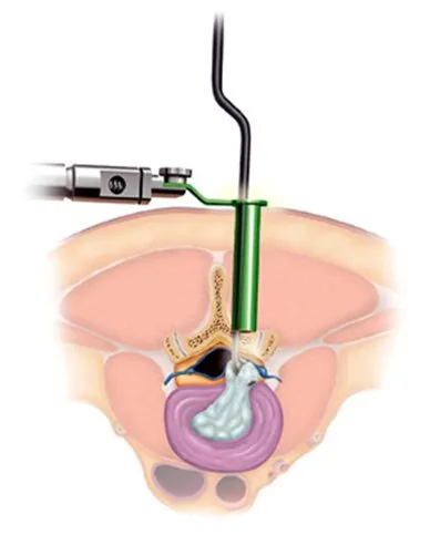 Enlarge image: Micro-endoscopic discectomy: the disc is reached through a narrow tube (illustration).
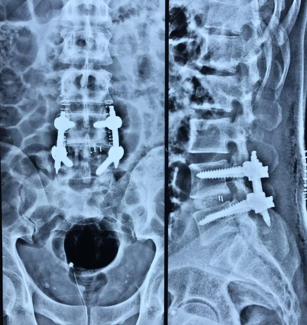 Enlarge image: X-rays after an L4-5 lumbar fusion with screws and rods.
Low-back conditions are extremely common, and many MRI changes are also seen in people who have little or no pain. Treatment therefore begins by matching symptoms and examination findings with imaging, rather than operating on a scan. Activity modification, structured physiotherapy, medicines and selected injections may be used first when there is no urgent neurological concern.
Surgery becomes more relevant when disabling leg symptoms persist despite appropriate conservative care, when instability or deformity is mechanically significant, or when progressive weakness, bladder/bowel dysfunction or other neurological warning signs make delay unsafe.
Care ranges from medicines and physiotherapy to targeted decompression when persistent nerve compression causes disabling symptoms.
Narrowing around the nerves can cause leg pain, heaviness or reduced walking tolerance and is treated according to severity and function.
When one vertebra slips relative to another, treatment depends on symptoms, nerve compression, instability and response to conservative care.
Kyphoplasty & Vertebroplasty
Fragility fractures need assessment of pain, collapse, alignment, nerve status and underlying bone health.
Typical goals of treatment
From Dr. Sharma's practice
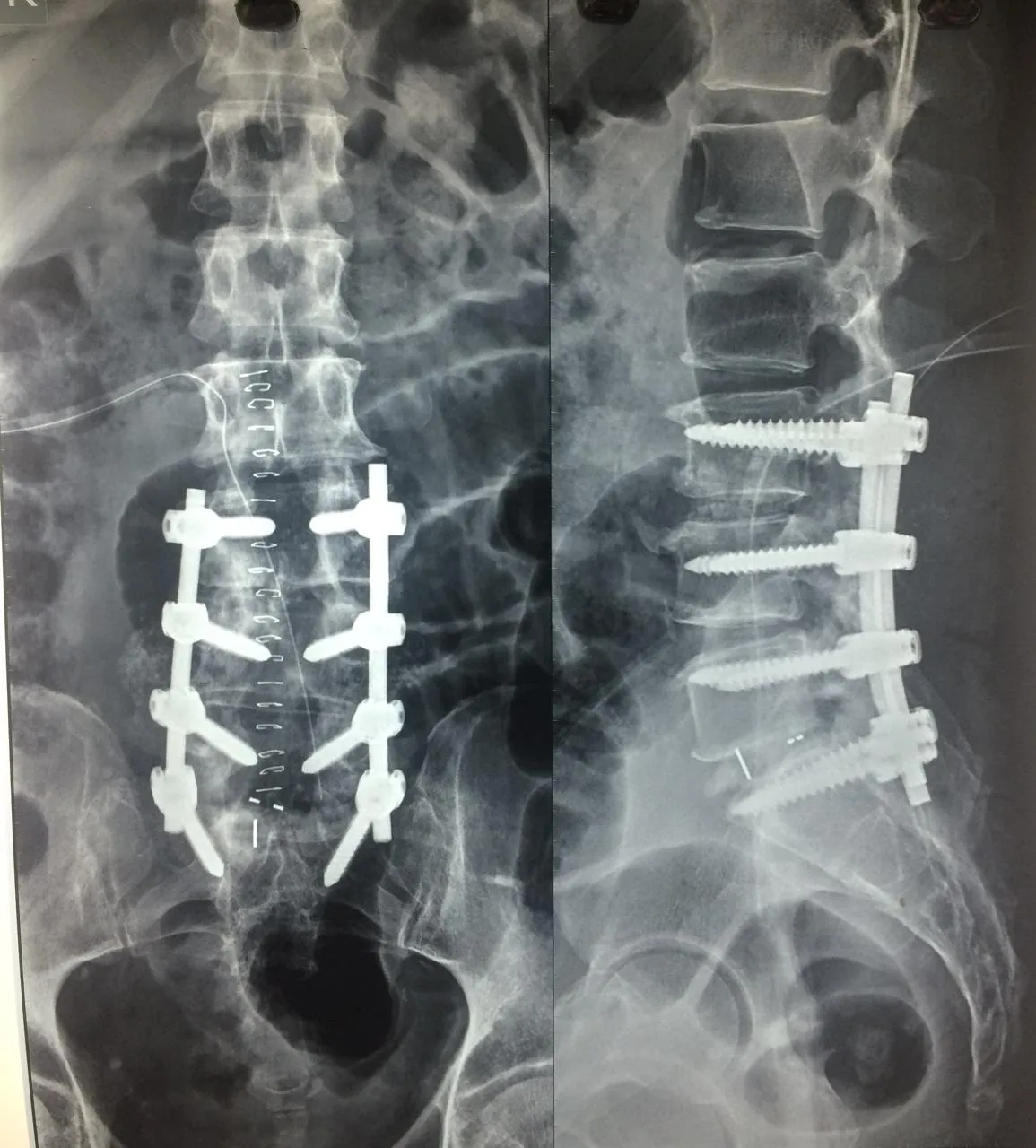 Enlarge image: X-rays after a multilevel lumbar spine fusion.
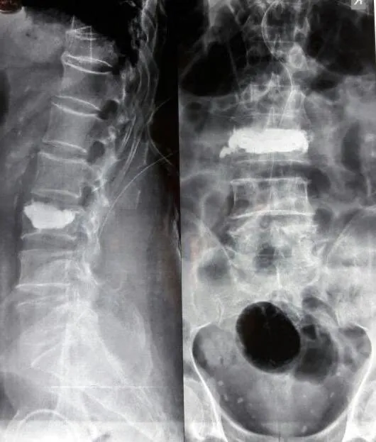 Enlarge image: Vertebroplasty: cement stabilising a fractured lumbar vertebra (post-op X-ray).
A compressed cervical nerve root usually produces pain, tingling, numbness or weakness in a particular arm distribution. Cervical myelopathy involves the spinal cord itself and may instead show up as clumsy hands, difficulty with buttons or handwriting, imbalance, heaviness in the legs or a change in walking pattern.
Treatment is matched to the structure causing compression and the neurological risk. Some nerve-root problems improve without surgery; progressive spinal-cord compression requires a different level of urgency because established neurological loss may not always fully reverse.
Disc and joint degeneration can irritate a nerve and produce neck-to-arm pain, tingling, numbness or weakness.
Cord compression may affect hand dexterity, balance and walking and needs timely specialist assessment.
Fusion can decompress neural structures and stabilize selected levels when movement or alignment is unsafe.
Motion-preserving disc replacement can be considered in appropriately selected cervical disc disease.
Procedure selection considers
From Dr. Sharma's practice
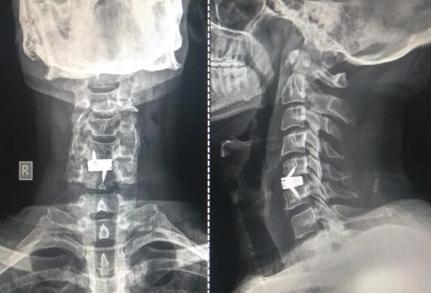 Enlarge image: Anterior cervical discectomy and fusion (ACDF), single level, stand-alone cage.
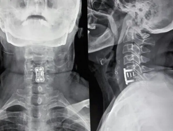 Enlarge image: ACDF with a cage and plate.
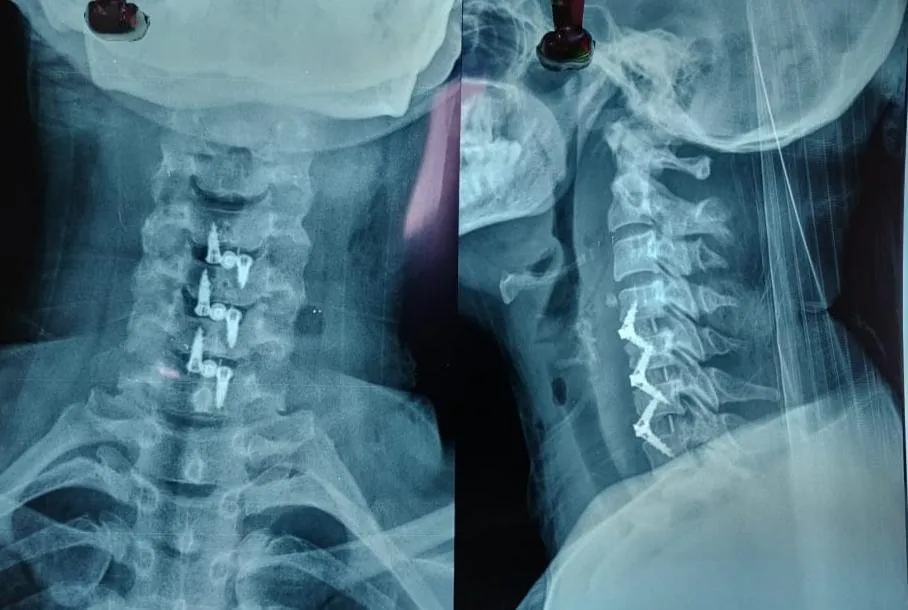 Enlarge image: Multilevel ACDF with stand-alone cages.
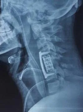 Enlarge image: Anterior cervical corpectomy and fusion.
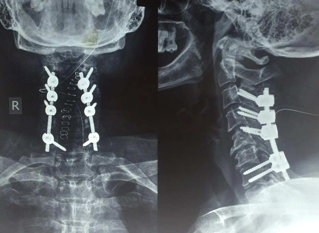 Enlarge image: Posterior cervical fusion with screws and rods.
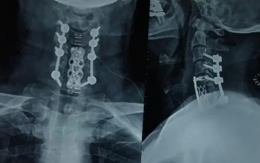 Enlarge image: Combined anterior and posterior cervical fusion.
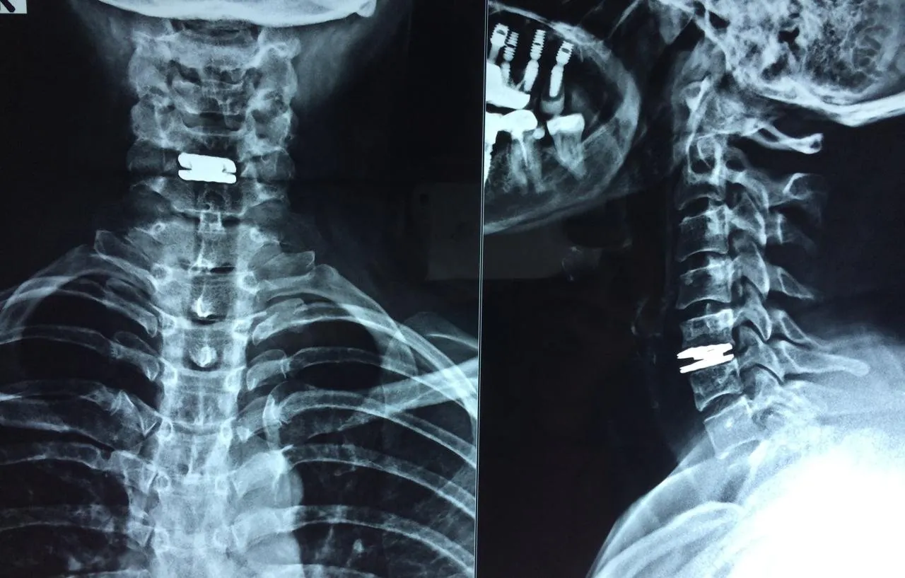 Enlarge image: Cervical disc replacement: an artificial disc that keeps the level moving.
Deformity, infection, tumor and revision surgery require more than a single-problem approach. The surgeon has to consider neurological compression, alignment, stability, bone quality, previous implants or scar tissue, infection control and the patient's overall medical reserve at the same time.
These cases may involve staged treatment, longer constructs, reconstruction or multidisciplinary input. The objective is not simply to make an X-ray look straighter; it is to improve or protect function while balancing the benefit of correction against the physiological cost of surgery.
Adult & Pediatric
Assessment considers curve size, progression, balance, pain, neurological function and the patient's age and goals.
Treatment combines microbiological diagnosis and prolonged medical therapy with surgery when instability, abscess, deformity or neural compression requires it.
Complex failed-back cases
Revision planning identifies the actual pain generator, residual compression, non-union, implant issues, adjacent-level disease or deformity before another operation is considered.
Management is individualized around tumor biology, neurological risk, structural stability and coordination with oncology.
Detailed evaluation can include
From Dr. Sharma's practice
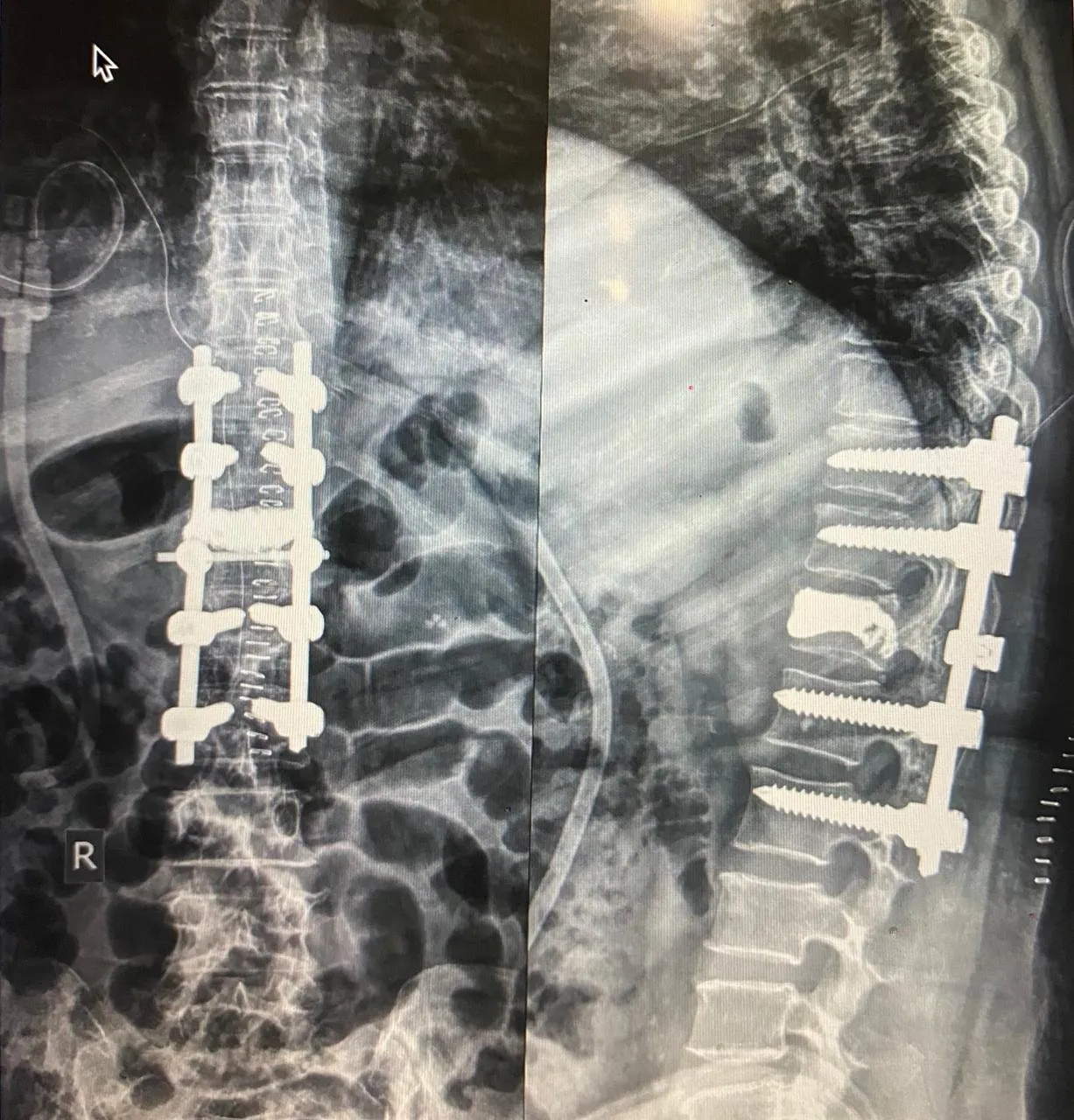 Enlarge image: Vertebroplasty combined with spinal instrumentation for a fracture in weak bone (post-op X-rays).
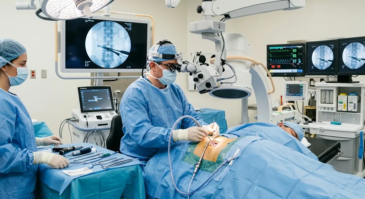 Minimally Invasive Spine SurgeryDecompression, stabilisation and fusion through small, muscle-sparing access corridors instead of a long open incision.Read more
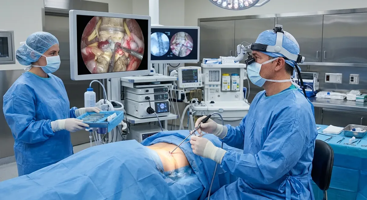 Endoscopic Spine SurgeryA thin camera and fine instruments pass through a very small incision to treat disc herniations and stenosis.Read more
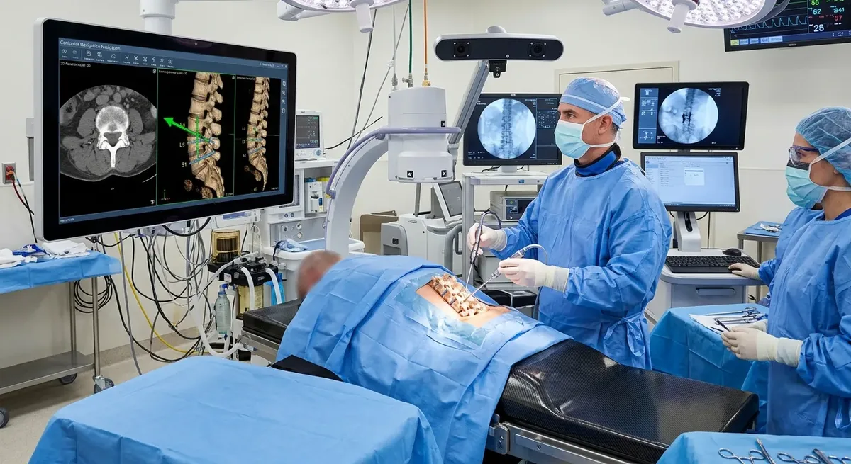 Computer Navigation Spine SurgeryShows the position of instruments relative to your spine on a 3D image in real time, guiding accurate screw and implant placement.Read more
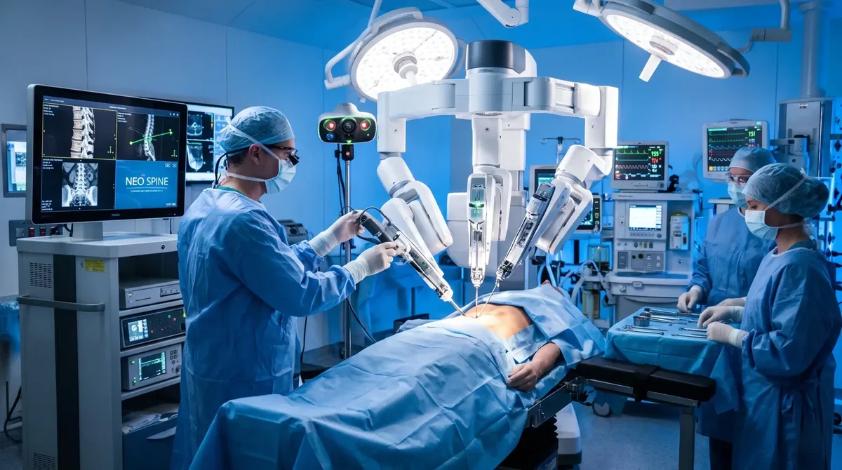 Robotic Spine SurgeryCT-based planning and a robotic guidance arm help the surgeon place spinal screws with 97 to 99 percent accuracy, even in complex anatomy.Read more
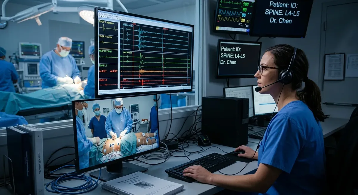 Spinal NeuromonitoringContinuous checks of spinal cord and nerve function during surgery, so the team is warned early if nerves are under stress.Read more
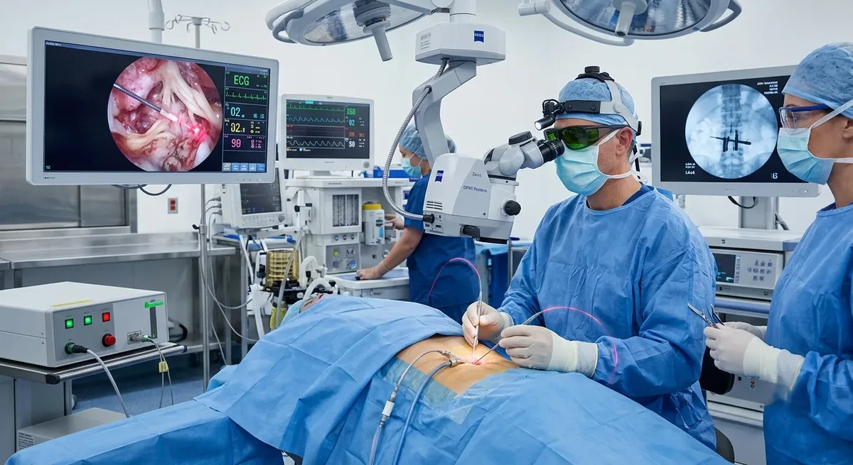 LASER Spine SurgeryFocused light energy used as a precise tool in selected minimally invasive procedures, often alongside endoscopic surgery.Read more
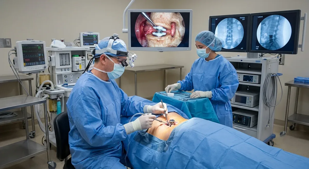 Disc Replacement SurgeryA worn disc is replaced with an artificial disc that keeps movement at that level, instead of fusing it.Read more
Ask Dr. Sharma. A consultation, or a review of your existing MRI, gives you a clear answer.
Treatment decisions depend on symptoms, examination findings, imaging and how much the problem is affecting function, not on an MRI label alone.
No. Many disc-related symptoms improve with time, activity modification, medication and physiotherapy. Surgery is generally considered when symptoms remain disabling despite appropriate non-surgical care, or sooner when there is significant or progressive neurological loss or another urgent warning sign.
The best approach is the one that safely and completely treats the actual pathology. A minimally invasive corridor can reduce tissue disruption in suitable cases, but a wider exposure may be safer or more effective for some deformities, complex revisions, infections or multi-level problems.
Fusion is used when the spine needs added stability, for example in selected cases of instability, spondylolisthesis, deformity, reconstruction or when decompression would otherwise leave a segment unstable. It is not automatically required for every decompression or disc operation.
The decision combines the symptom pattern, neurological examination, imaging, spinal alignment and stability, previous treatment, age, bone quality, medical health and the patient's priorities. The operation should have a specific anatomical and functional goal that can be clearly explained before surgery.
Send your scans and reports securely for Dr. Sharma to review, or book a consultation to discuss them in person.