What Is Spondylolisthesis?
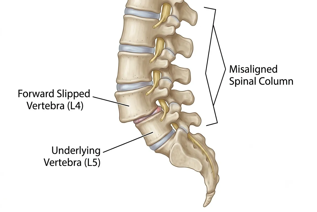
Spondylolisthesis is a spinal condition in which one vertebra slips forward relative to the vertebra immediately below it. The name comes from terms referring to the spine and slipping or sliding. In adults, the condition is often linked to degeneration of the discs and facet joints that normally stabilize a spinal segment.
When these stabilizing structures deteriorate, abnormal movement can develop between adjacent vertebrae. Spondylolisthesis can also be associated with spondylolysis—a defect or fracture through the pars interarticularis in the posterior part of a vertebra. A pars defect disrupts part of the bony mechanism that helps keep the upper vertebra aligned over the one below.
The amount of slip is only part of the diagnosis
Treatment depends on symptoms, neurological compression and whether the segment is unstable—not simply on the grade seen on an X-ray.
Affected Regions and Grades
Spondylolisthesis occurs most commonly in the lumbar spine, although vertebral slippage can also occur in the cervical spine. The degree of displacement is commonly described by the percentage of one vertebral body that has translated over the next.
| Grade | Approximate Slip | Clinical Meaning |
|---|---|---|
| Grade I | Up to 25% | Low-grade slip; many patients can be managed non-surgically when symptoms are mild and the segment is stable. |
| Grade II | 26–50% | Moderate displacement; symptoms, stenosis and dynamic instability become increasingly important in treatment planning. |
| Grade III | 51–75% | High-grade slip requiring careful assessment for progression, deformity and neurological compromise. |
| Grade IV | 76–100% | Severe displacement; surgical stabilization is more frequently considered when clinically appropriate. |
Higher-grade slips are more likely to be associated with significant symptoms or require advanced treatment, but a lower-grade slip can still be important if it is unstable or causes substantial nerve compression.
Spondylolisthesis, Spondylolysis and Spondylosis
| Term | What It Means | Typical Imaging Finding |
|---|---|---|
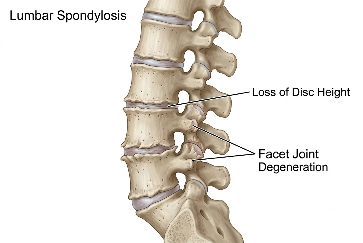
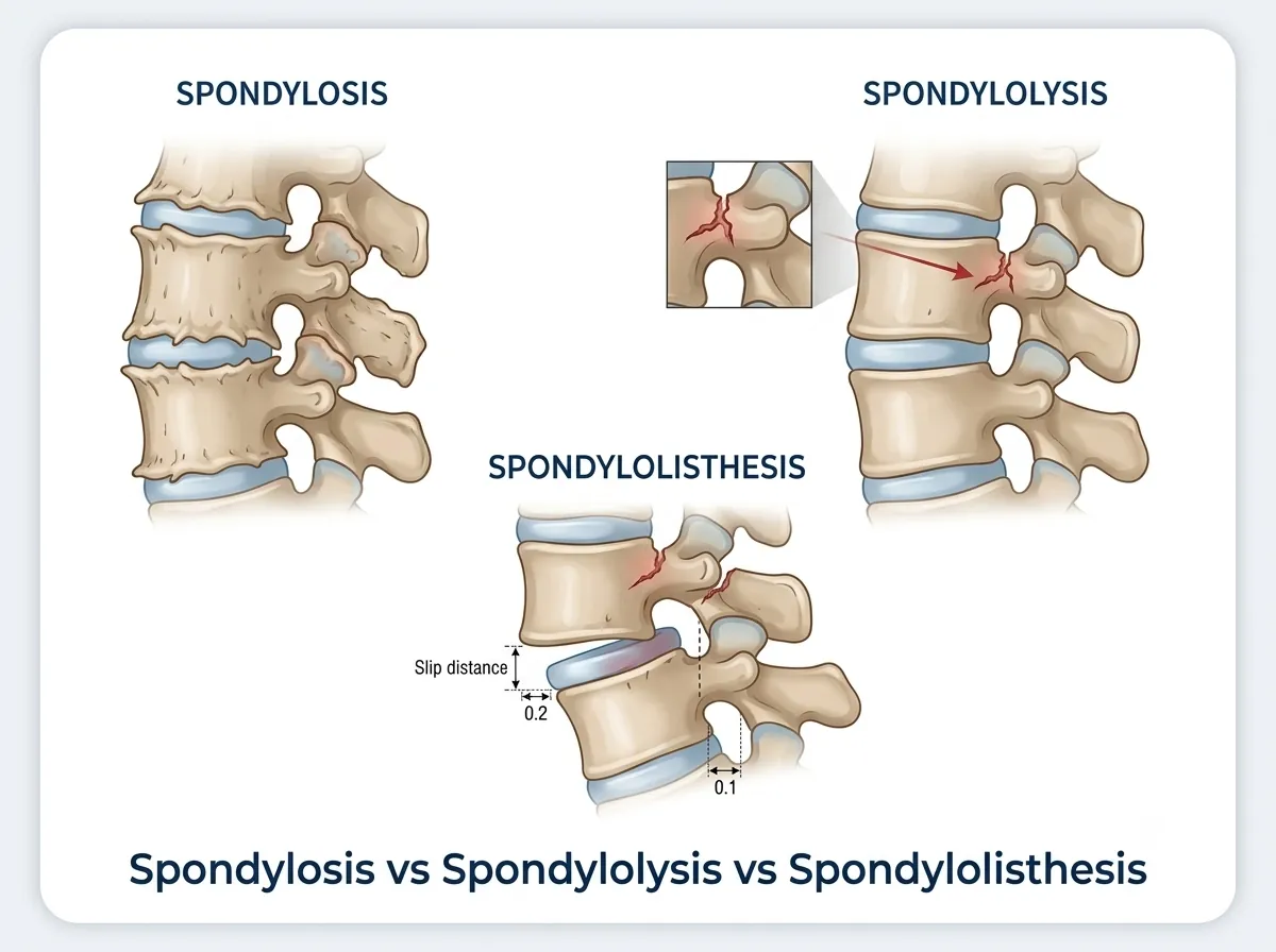
| Spondylosis | Degenerative wear of spinal discs, facet joints and adjacent bone. | Disc-space loss, osteophytes and facet-joint degeneration. |
| Spondylolysis | A defect or fracture through the pars interarticularis. | Pars defect visible on appropriate X-ray or CT imaging. |
| Spondylolisthesis | Translation of one vertebra relative to the vertebra below. | Visible vertebral slippage, which can be measured and graded on standing radiographs. |
These conditions can coexist. Degenerative spondylosis can weaken segmental stability and contribute to a slip, while a pars defect can permit one vertebra to translate forward. Ankylosing spondylitis is different: it is an inflammatory disease rather than a vertebral slippage disorder.
Symptoms
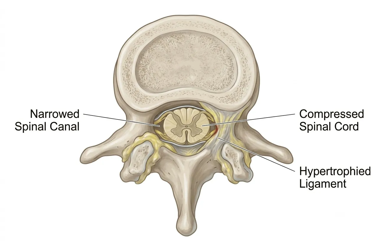
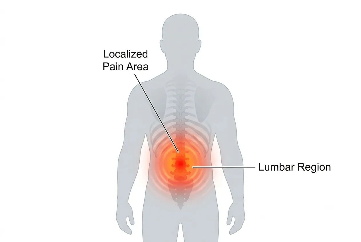
The clinical picture depends on the location, severity and duration of the slip. Localized back pain related to associated degenerative change is common. Spondylolisthesis can also disturb the normal dimensions and alignment of the spinal canal, and degenerative changes may add further stenosis.
Symptoms When Nerves or the Spinal Cord Are Compressed
- Radiating pain into an arm or leg, depending on the spinal level involved.
- Tingling, numbness or reduced sensation.
- Muscle weakness.
- Reduced hand grip when cervical neural structures are affected.
- Loss of coordination or increasing difficulty with balance and walking.
- Abnormal limb tightness or stiffness when the spinal cord is involved.
- Bladder or bowel dysfunction in severe neurological compression.
Seek urgent assessment
New bladder or bowel dysfunction, rapidly progressive weakness, severe loss of coordination or major neurological deterioration needs prompt evaluation.
Arrange specialist review
Increasing radiating pain, numbness, weakness, reduced walking tolerance or persistent disabling back pain should be assessed.
Routine conservative care
Mild stable symptoms without neurological deficit can often begin with structured non-surgical management and follow-up.
2-minute self-check
Do your symptoms deserve a closer look?
Tick anything you've noticed recently. Your guidance appears straight below. Everything stays on this page; nothing is stored or sent. This is an educational aid, not a diagnosis of spondylolisthesis.
Seek urgent medical care today
These symptoms can mean a nerve or the spinal cord is under serious pressure. Go to the nearest emergency department, or call the clinic straight away. Do not wait for a routine appointment.
Book a spine specialist review soon
Arrange an assessment within the next few days, and sooner if things get worse. If you already have an MRI, you can send it for review.
Conservative care is usually a reasonable start
Activity changes, physiotherapy and appropriate medication often help. See a doctor if it is not better in 1–2 weeks, or straight away if any time-sensitive symptom appears.
Guidance only, based on the warning signs described in this guide. It cannot find the cause of your symptoms. If symptoms are severe, getting worse quickly, or you are worried, contact a doctor.
Diagnosis
Assessment establishes the amount of vertebral slippage, looks for instability, identifies a pars defect when present and determines whether stenosis or neural compression explains the patient's symptoms.
Imaging and Diagnostic Tests
- X-ray: demonstrates the slip and allows grading. It can also show disc-height loss, osteophytes and other signs of spondylosis.
- Flexion-extension X-rays: compare alignment in different positions and can reveal dynamic instability that is not apparent on a single static image.
- MRI: assesses the slip together with discs, stenosis, nerve roots and, where relevant, the spinal cord.
- CT: provides detailed bony anatomy and is particularly useful for confirming or excluding a pars interarticularis defect.
- EMG/NCV: may help assess nerve function when neurological symptoms require further clarification.
- Blood tests: are used selectively to exclude associated medical conditions when clinically indicated.
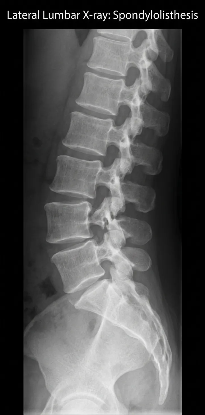 Enlarge image: X-ray showing vertebral slippage in spondylolisthesis
Non-Surgical Treatment
Treatment is individualized according to symptoms, slip grade, neurological findings and segmental stability. Patients with mild symptoms and a lower-grade stable slip often respond well to conservative care.
- Pain medication: appropriate analgesic or anti-inflammatory treatment may be used according to the patient's medical profile.
- Heat or ice: local application may provide short-term symptom relief.
- Activity modification: temporarily restricting movements or loads that repeatedly provoke pain can help settle an acute episode.
- Bracing: selected patients may use temporary external support when clinically appropriate.
- Physiotherapy and exercise: strengthening the core and spinal muscles creates an effective muscular support system and can reduce excessive stress across the affected segment.
Pain Management Procedures
Interventional Pain Options
- Intradiscal steroid injections in selected disc-related pain patterns.
- Laser disc therapy in carefully selected cases.
- Facet-joint injections when the facet joint is suspected to be a pain generator.
- Radiofrequency ablation for selected facet-mediated pain.
- Epidural steroid injections when nerve-root inflammation or compression contributes to radiating symptoms.
Pain procedures do not correct the slip
Injections and other interventional procedures may reduce symptoms in selected patients, but they do not reverse vertebral displacement, a pars defect or established degenerative change.
Surgical Treatment
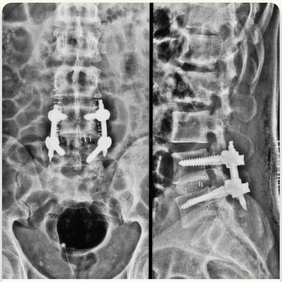
Surgery is considered when adequate conservative treatment fails, symptoms are severe or neurological compression and instability require structural correction. The operation should address the actual problem rather than adding fusion automatically to every case.
| Surgical Strategy | When It May Be Considered |
|---|---|
| Decompression without fusion | Selected lower-grade slips where neural compression is the dominant problem, back pain is limited and clinically significant instability is absent. |
| Fusion with instrumentation | A pars defect, high-grade slip, significant mechanical back pain from degeneration, demonstrated instability, associated deformity or situations where adequate decompression requires extensive removal of stabilizing bone or joints. |
When Fusion May Not Be Appropriate
- Severe osteoporosis: poor bone quality can compromise implant fixation and delay or reduce the reliability of bone fusion.
- Major medical comorbidity: serious health conditions can make the added magnitude of fusion surgery inappropriate for some patients.
- Compression without a fusion indication: if neural decompression is required but the segment does not otherwise need stabilization, unnecessary fusion can add complexity, cost and recovery burden.
Minimally Invasive Surgery
Many operations for spondylolisthesis can now be performed using minimally invasive or keyhole techniques when the anatomy and surgical objective are suitable. The aim is to achieve the required decompression and/or stabilization while reducing disruption of surrounding tissues.
- Reduced blood loss compared with larger open exposures in suitable cases.
- Less postoperative tissue pain and potentially lower analgesic requirements.
- Earlier mobilization and rehabilitation.
- Shorter hospital stay for appropriately selected patients.
- Smaller surgical scars.
The technique is chosen according to the slip, stenosis, deformity, bone quality and required reconstruction. Minimally invasive surgery is a method of access; the essential goal remains adequate nerve decompression and reliable stabilization when fusion is required.
Imaging from Dr. Sharma's practice
The images below are from Dr. Amit Sharma's practice and are shared for education, without identifying details. Every patient's imaging is different — your own scans need an individual clinical review.
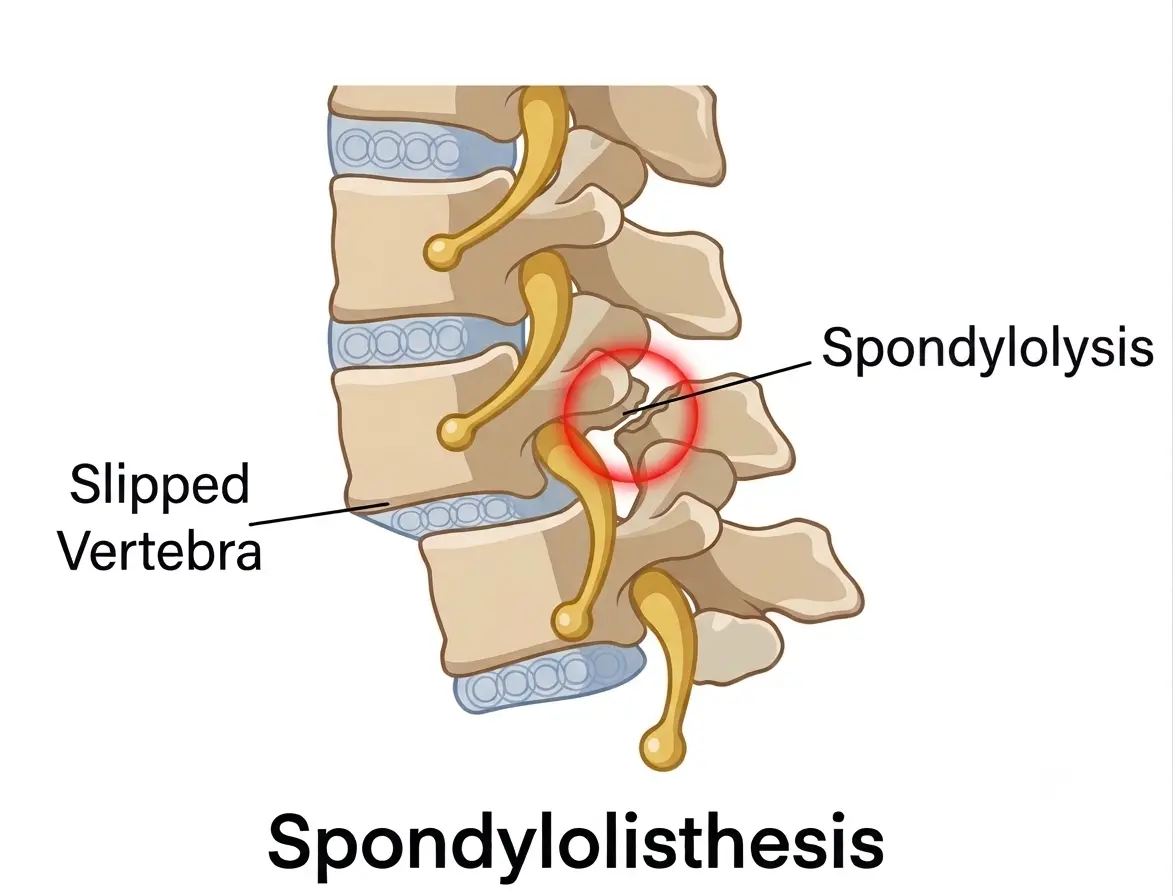 Enlarge image: Illustration of spondylolysis (a defect in the pars) and the resulting slipped vertebra.
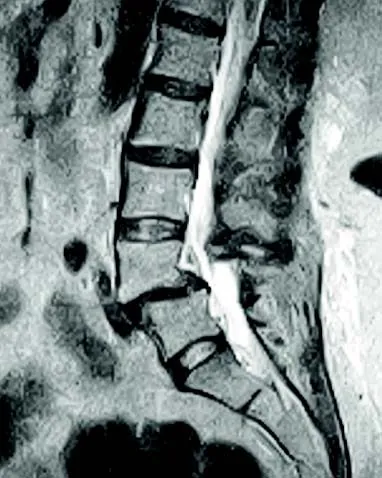 Enlarge image: Sagittal lumbar MRI showing a vertebra slipped forward on the one below.
Frequently Asked Questions
What is the difference between spondylolisthesis and spondylosis?
Spondylolisthesis means one vertebra has slipped relative to another. Spondylosis describes degenerative wear of discs and facet joints that can cause stiffness and pain.
How is spondylolysis different?
Spondylolysis is a defect or fracture through the pars interarticularis. It may exist without vertebral slippage or may permit a vertebra to slip, producing isthmic spondylolisthesis.
What factors can make spondylolisthesis worse?
Progressive degeneration, repeated mechanical loading, instability, a pars defect and high-grade displacement can increase symptom burden or the risk of progression in selected patients.
Can spondylolisthesis be treated without surgery?
Yes. Mild, lower-grade and stable slips often respond to medication, activity modification, physiotherapy, core strengthening and other conservative measures.
How does ankylosing spondylitis differ from spondylolisthesis?
Ankylosing spondylitis is an inflammatory disease affecting the spine and sacroiliac joints. Spondylolisthesis is a mechanical or structural vertebral slip.
What does an X-ray show in spondylosis and spondylolisthesis?
Spondylosis may appear as disc-space narrowing, osteophytes and facet degeneration. Spondylolisthesis appears as measurable displacement of one vertebra relative to the next; dynamic X-rays can additionally demonstrate instability.
How do symptoms of spondylolisthesis and spondylolysis differ?
A pars defect can cause mechanical back pain and may be asymptomatic. Once slippage or associated stenosis develops, spondylolisthesis can additionally produce nerve-related pain, numbness, weakness or walking difficulty.
What are the radiological differences among spondylosis, spondylolysis and spondylolisthesis?
Spondylosis is identified by degenerative disc and joint changes; spondylolysis by a pars interarticularis defect; and spondylolisthesis by translation of one vertebra over another. CT is particularly useful for pars anatomy, while standing and flexion-extension radiographs assess the slip and instability.
This article is general education written by Dr. Amit Sharma. It is not a diagnosis or a substitute for an in-person consultation. If you have sudden weakness, numbness in the groin or saddle area, or new bladder or bowel problems, seek emergency care.