Understanding Scoliosis
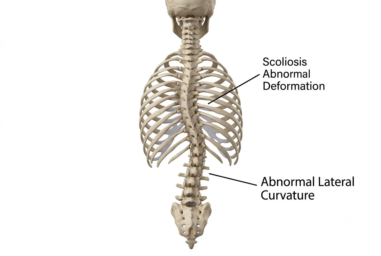
When viewed from behind, a healthy spine is broadly straight. Scoliosis is a sideways spinal curvature, often accompanied by vertebral rotation. The curve may be C-shaped or form a double S-shaped pattern.
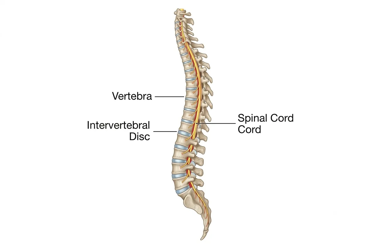
| Location | Region Involved |
|---|---|
| Cervical | Neck region. |
| Thoracic | Upper and middle back. |
| Lumbar | Lower back. |
Causes and Types
Major Types of Scoliosis
- Idiopathic: the commonest group; the precise cause is unknown. It occurs more often in girls and can have a familial/genetic component.
- Congenital: abnormal vertebral development produces an often sharp curve that may progress rapidly.
- Neuromuscular: poor muscular support or abnormal neurological control produces long curves, often C-shaped.
- Degenerative: asymmetric wear of adult spinal segments gradually creates curvature.
- Secondary: uneven bone destruction from infection, fracture or tumor can create a deformity.
- Syndromic: scoliosis can accompany systemic disorders including Marfan syndrome, Down syndrome, osteogenesis imperfecta and neurofibromatosis.
Apparent or functional scoliotic curvature may also arise from unequal leg length with pelvic obliquity, muscle spasm or disc-related pain.
Classification by Age
| Group | Age | Why It Matters |
|---|---|---|
| Early onset | Under 10 years | Large remaining growth potential means progression can affect chest and lung development. |
| Late onset | Over 10 years until skeletal maturity | Remaining growth and curve magnitude guide observation, bracing and surgical timing. |
| Adult onset | After skeletal maturity | Degeneration, pain, stenosis and balance become major treatment considerations. |
Symptoms
- Visible spinal curvature and cosmetic asymmetry.
- Rib hump or asymmetric lumbar muscle prominence.
- Unequal shoulder height, waist asymmetry or uneven chest prominence.
- Trunk shift or body tilt to one side.
- Difficulty sitting, standing or walking in severe or neuromuscular deformity.
- Back pain, particularly in degenerative scoliosis; muscle imbalance can also produce discomfort in idiopathic curves.
Severe deformity can narrow the spinal canal and produce weakness, numbness, impaired coordination, spasticity or bladder/bowel dysfunction. Congenital scoliosis may coexist with intraspinal abnormalities such as tethered cord or diastematomyelia, and congenital anomalies may also involve the heart, kidneys or abdomen. Very early severe curves can impair chest and lung growth and, in extreme cases, breathing.
Urgent
New weakness, major sensory loss, severe balance deterioration or bladder/bowel dysfunction requires prompt assessment.
Specialist review
A visibly progressing curve, increasing imbalance, significant pain or a rapidly changing childhood deformity should be reviewed.
Observation
Mild stable curves can often be monitored, but follow-up is important while significant growth remains.
2-minute self-check
Do your symptoms deserve a closer look?
Tick anything you've noticed recently. Your guidance appears straight below. Everything stays on this page; nothing is stored or sent. This is an educational aid, not a diagnosis of scoliosis.
Seek urgent medical care today
These symptoms can mean a nerve or the spinal cord is under serious pressure. Go to the nearest emergency department, or call the clinic straight away. Do not wait for a routine appointment.
Book a spine specialist review soon
Arrange an assessment within the next few days, and sooner if things get worse. If you already have an MRI, you can send it for review.
Conservative care is usually a reasonable start
Activity changes, physiotherapy and appropriate medication often help. See a doctor if it is not better in 1–2 weeks, or straight away if any time-sensitive symptom appears.
Guidance only, based on the warning signs described in this guide. It cannot find the cause of your symptoms. If symptoms are severe, getting worse quickly, or you are worried, contact a doctor.
Diagnosis
Evaluation defines the magnitude and cause of the deformity, the patient's main concern, neurological status and remaining growth. Examination includes overall balance, curve pattern, neurological findings and signs of puberty/skeletal maturity, plus features suggesting congenital, neuromuscular or secondary disease.
Diagnostic Tests
- Standing X-rays: measure curve magnitude, including Cobb angle, and permit interval comparison during observation.
- Bending X-rays: demonstrate curve flexibility and assist surgical planning.
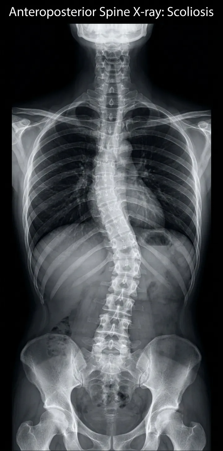 Enlarge image: Scoliosis X-ray showing spinal curvature
MRI, CT and Additional Tests
- MRI: evaluates the spinal cord, congenital anomalies, stenosis and degenerative or secondary pathology when clinically indicated.
- CT/3D reconstruction: defines complex bone anatomy and can aid deformity and instrumentation planning.
- EMG/NCV: may assess abnormal nerve function.
- Additional tests: abdominal ultrasound and echocardiography may investigate associated congenital anomalies; biopsy may be required when infection or tumor is suspected.
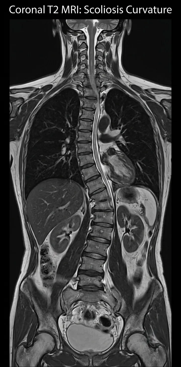 Enlarge image: Scoliosis MRI showing the spinal canal and neural structures
Non-Surgical Treatment
Treatment depends on age, curve type, magnitude, progression, symptoms and remaining growth. The three broad strategies are observation, bracing/casting and surgery.
| Strategy | Typical Role |
|---|---|
| Observation | Mild curves without major cosmetic, neurological or functional problems, or when definitive treatment is not presently appropriate. Growing children and higher-risk curve types require close surveillance. |
| Casting / bracing | Selected growing patients with borderline curves, commonly around 20–40°, to reduce progression and delay or avoid surgery. Casting may be used in very young children. |
| Symptom treatment | Physiotherapy, medication or injections can address pain and function but do not reliably straighten a structural scoliosis. |
Bracing does not reverse an established structural curve and is ineffective after skeletal maturity. Curves that progress despite bracing require reassessment. Braces may need replacement as a child grows, and congenital, neuromuscular and degenerative curves generally do not respond to bracing in the same way as adolescent idiopathic scoliosis.
Scoliosis Surgery
Surgery is considered for large or progressive curves, significant cosmetic deformity or imbalance, neurological risk, and curve types with a high likelihood of progression. Curves exceeding roughly 40–50° commonly enter the surgical discussion because larger, stiffer deformities become more difficult to correct safely.
Core Surgical Principles
- Correct the deformity as safely as possible while preventing further progression.
- Posterior instrumentation and fusion is the commonest approach for many idiopathic curves.
- Fusion levels are selected carefully because fused segments permanently lose motion.
- Selected rigid deformities may require anterior release or combined approaches.
- Motion-preserving/growth-friendly techniques can be considered in carefully selected flexible or growing spines, but they are not suitable for every curve.
Special Scoliosis Groups
- Early-onset scoliosis: growth-friendly, non-fusion constructs may control the curve while allowing chest and lung development before later definitive fusion.
- Congenital scoliosis: often progresses rapidly and may require early surgery; sharp curves and associated cord anomalies increase neurological complexity.
- Neuromuscular scoliosis: can progress rapidly and is often accompanied by sitting imbalance and nutritional or general medical challenges.
- Degenerative scoliosis: treatment must address both deformity and age-related stenosis/instability. Large reconstructive operations carry greater complication and implant-failure risk, particularly with osteoporosis and medical comorbidity. In selected patients, a smaller targeted decompression or short fusion may provide better risk-benefit balance than full deformity correction.
Risks and Modern Advances
| Issue / Advance | Clinical Relevance |
|---|---|
| Incomplete correction | Very large curves may only be safely partially corrected; complete correction is not required for a worthwhile cosmetic and functional result. |
| Neurological risk | Risk varies with age, cause, curve magnitude and correction attempted. Severe and congenital curves are higher risk. |
| Implant problems | Prominence, loosening, breakage, non-union or malposition can occur, with higher mechanical stress in non-fusion constructs. |
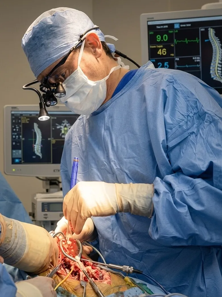
| Navigation / robotics | Provides intra-operative guidance for screw placement in rotated, deformed and small vertebrae. |
| Neuromonitoring | Continuously tracks spinal-cord and nerve function so concerning changes can be recognized during correction. |
| Blood conservation | Modern strategies including cell-saver technology can reduce reliance on donor transfusion in major deformity surgery. |
Recovery
Mobilization after modern scoliosis surgery can begin early; many patients are permitted to sit and walk from the following day as pain and medical condition allow. Advanced multimodal pain-management protocols support earlier rehabilitation. Overall recovery depends on curve severity, procedure extent, age and associated medical conditions.
The goal is safe correction, not a perfectly straight X-ray
For severe curves, forcing complete correction can increase neurological and mechanical risk. Surgery aims for a balanced, durable and safe correction while preserving as many mobile segments as practical.
Imaging from Dr. Sharma's practice
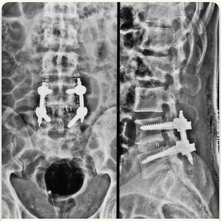
The images below are from Dr. Amit Sharma's practice and are shared for education, without identifying details. Every patient's imaging is different — your own scans need an individual clinical review.
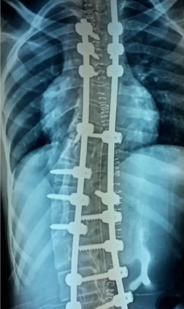 Enlarge image: Scoliosis: instrumented correction (clinical image).
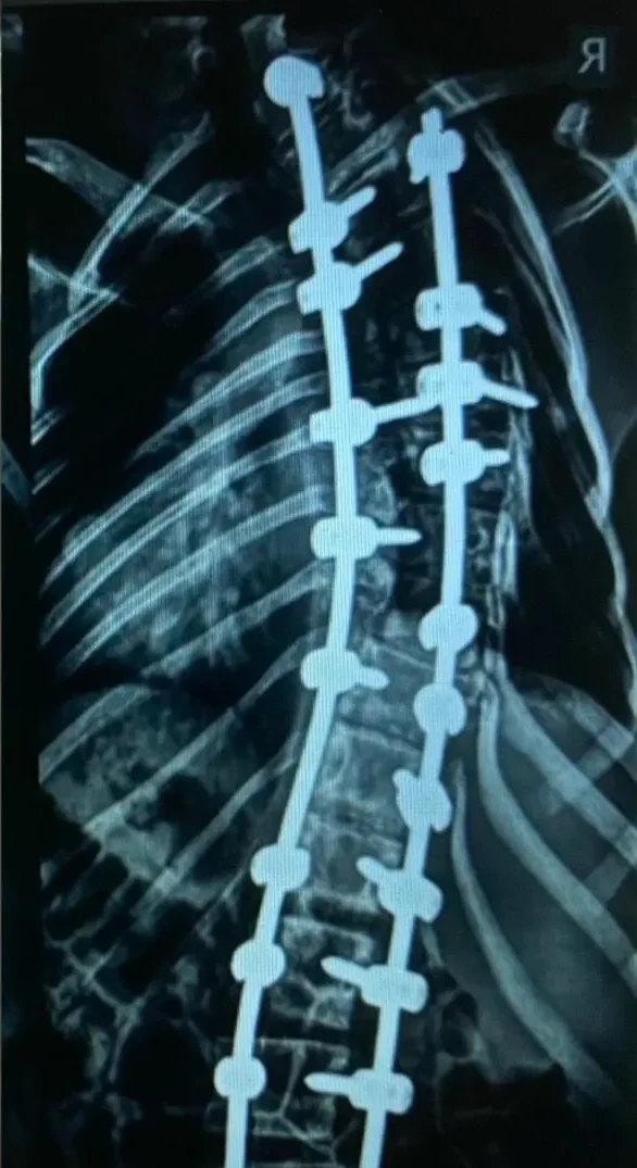 Enlarge image: Scoliosis: instrumented correction, second case (clinical image).
Frequently Asked Questions
Can scoliosis be cured by exercise?
Exercise can improve conditioning, posture and pain, but it does not reliably correct a significant structural curve. Scoliosis-specific therapy may support function as part of a broader treatment plan.
Are there scoliosis medicines?
Medication can treat pain or associated inflammation but cannot straighten the structural deformity.
What treatments are available without surgery?
Observation, periodic X-rays, casting in selected young children, bracing in selected growing patients, and symptom-directed physiotherapy or pain treatment.
Can adult scoliosis be corrected?
Yes, but treatment ranges from symptom-focused care to decompression, limited fusion or full deformity reconstruction depending on pain, stenosis, balance, curve severity and medical fitness.
What is the best age for surgery?
There is no single age. Timing depends on curve type, magnitude, progression and remaining growth; congenital and rapidly progressive curves may need earlier intervention.
How are teenagers treated?
Growing adolescents with mild curves may be observed, selected 20–40° curves may be braced, and larger or progressive curves may require surgery.
How is lumbar scoliosis treated?
Treatment depends on age and cause. Adult lumbar curves often require management of degenerative pain, stenosis and instability as well as the deformity itself.
What are mild symptoms?
Subtle shoulder or waist asymmetry, a small rib prominence, mild trunk shift or little/no pain may be the first signs.
What does scoliosis pain feel like?
Pain is commonly mechanical or muscular; adults may also have nerve-related leg pain from degenerative stenosis.
Is scoliosis curable?
Some curves remain mild and need only observation. Significant structural curves are managed rather than “cured” by exercise; surgery can substantially correct and stabilize severe deformity.
Can scoliosis be prevented?
Most idiopathic and congenital scoliosis cannot be prevented. Early detection and monitoring can, however, identify progression while less invasive options remain possible.
What newer treatments exist for adults?
Modern care includes navigation, robotic guidance, neuromonitoring, improved implants, minimally disruptive approaches in selected cases and individualized limited versus full reconstruction.
How long is surgical recovery?
Walking often starts early, but return to normal activity develops over weeks to months depending on the magnitude of surgery and individual healing.
What does scoliosis surgery cost?
Cost varies substantially with curve complexity, number of levels, implants, technology used and hospital stay; a meaningful estimate requires individual surgical planning.
What is the success rate?
Outcome depends on the curve and goals of treatment. Modern surgery can provide durable correction and stabilization, but no percentage applies to every scoliosis type.
What are the main surgical risks?
Neurological injury, infection, bleeding, inadequate correction, implant problems, non-union and general surgical complications are considered during consent and planning.
This article is general education written by Dr. Amit Sharma. It is not a diagnosis or a substitute for an in-person consultation. If you have sudden weakness, numbness in the groin or saddle area, or new bladder or bowel problems, seek emergency care.