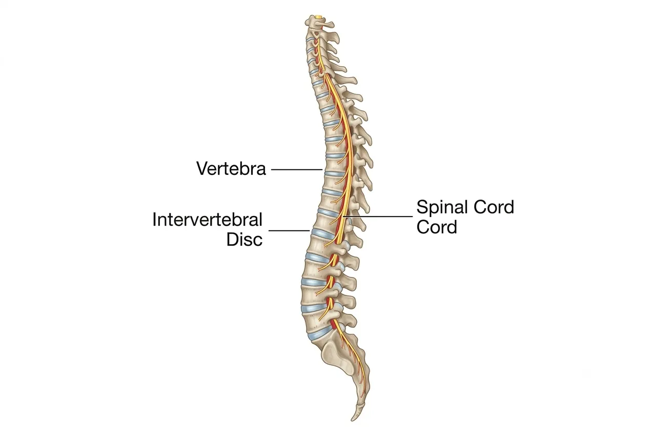
The human spine, or backbone, extends from the base of the skull to the pelvis and forms the central structural support of the body. It keeps the head and trunk balanced, supports upright posture, permits bending and rotation, and protects the spinal cord as it travels through the spinal canal.
Normal Spine Structure
A normal spine is a carefully balanced column of bones, joints, discs, ligaments, muscles and nerves. The vertebrae are stacked one above another, with intervertebral discs positioned between most vertebral bodies. Together, these structures provide both stability and controlled flexibility rather than functioning as a rigid column.
The spinal column also surrounds and protects the spinal cord and contributes to protection of important internal structures. Its shape and arrangement allow the body to tolerate everyday forces generated by standing, walking, lifting and other movements.
Natural Curvature of the Spine
Viewed from the side, a healthy spine is not straight. It has a characteristic gentle “S” profile created by three principal curves. The cervical and lumbar regions curve forward, producing lordosis, while the thoracic region curves backward, producing kyphosis.
These normal curves help keep the head and trunk aligned over the pelvis. They also distribute body weight, improve balance, absorb shock and reduce the concentration of mechanical stress on individual vertebrae and discs.
Regions of the Spine
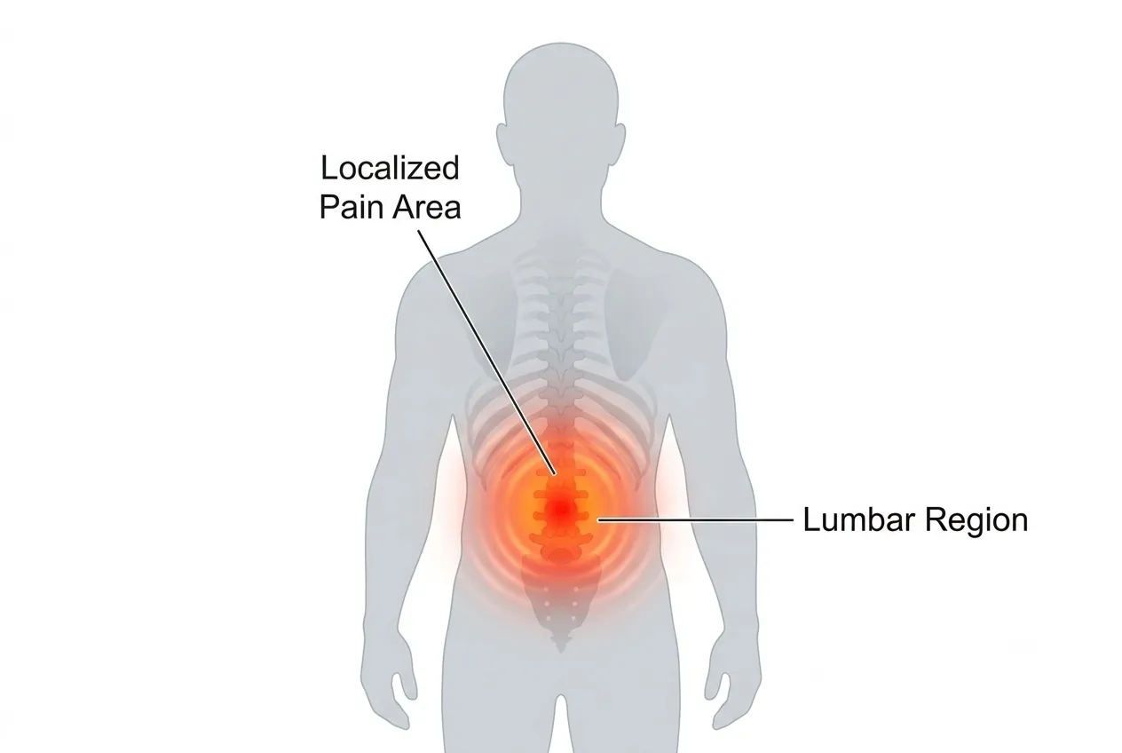
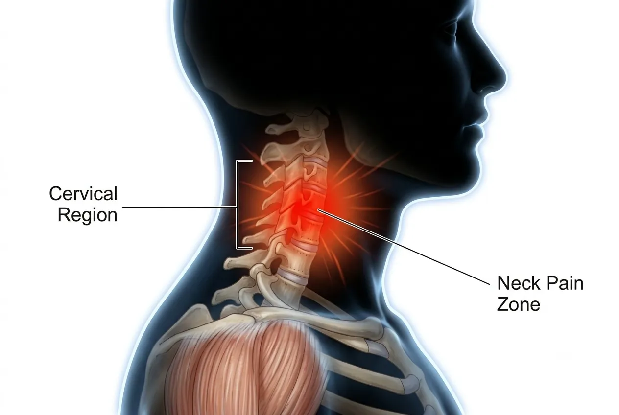
The spinal column is divided into the cervical, thoracic and lumbar regions, followed by the sacrum and coccyx. Each region has a different anatomical design because it performs a different combination of movement, stability, protection and weight-bearing.
Cervical Spine — Neck Region
- The cervical spine contains seven vertebrae, C1 to C7.
- It supports the head while allowing a large range of neck movement.
- Its normal forward curve is called cervical lordosis.
- C1 (Atlas) and C2 (Axis) are specially shaped upper cervical vertebrae that contribute substantially to nodding, rotation and tilting of the head.
Thoracic Spine — Upper and Middle Back
- The thoracic spine contains 12 vertebrae, T1 to T12.
- It forms the upper and middle back and provides attachment points for the ribs.
- The ribs and thoracic vertebrae contribute to the rib cage, helping protect organs including the heart and lungs.
- Its normal backward curve is called thoracic kyphosis.
- Compared with the neck and lower back, this region is designed more for stability and protection and therefore has relatively less mobility.
Lumbar Spine — Lower Back
- The lumbar spine usually contains five vertebrae, L1 to L5.
- Normal anatomical variation can occur, and some people may have four or six lumbar-type vertebrae.
- The lumbar spine connects the thoracic spine to the pelvis and carries a substantial proportion of body weight.
- Its normal forward curve is called lumbar lordosis.
- This region combines weight-bearing strength with the mobility required for bending and everyday trunk movement.
Sacrum and Coccyx
- The sacrum is a triangular bone below the lumbar spine formed by vertebrae that fuse during development.
- It connects the spinal column with the pelvis and plays an important role in transferring load and providing stability.
- The coccyx, or tailbone, lies below the sacrum and consists of a small group of fused bones.
- The coccyx provides attachment for several muscles and ligaments in the pelvic region.
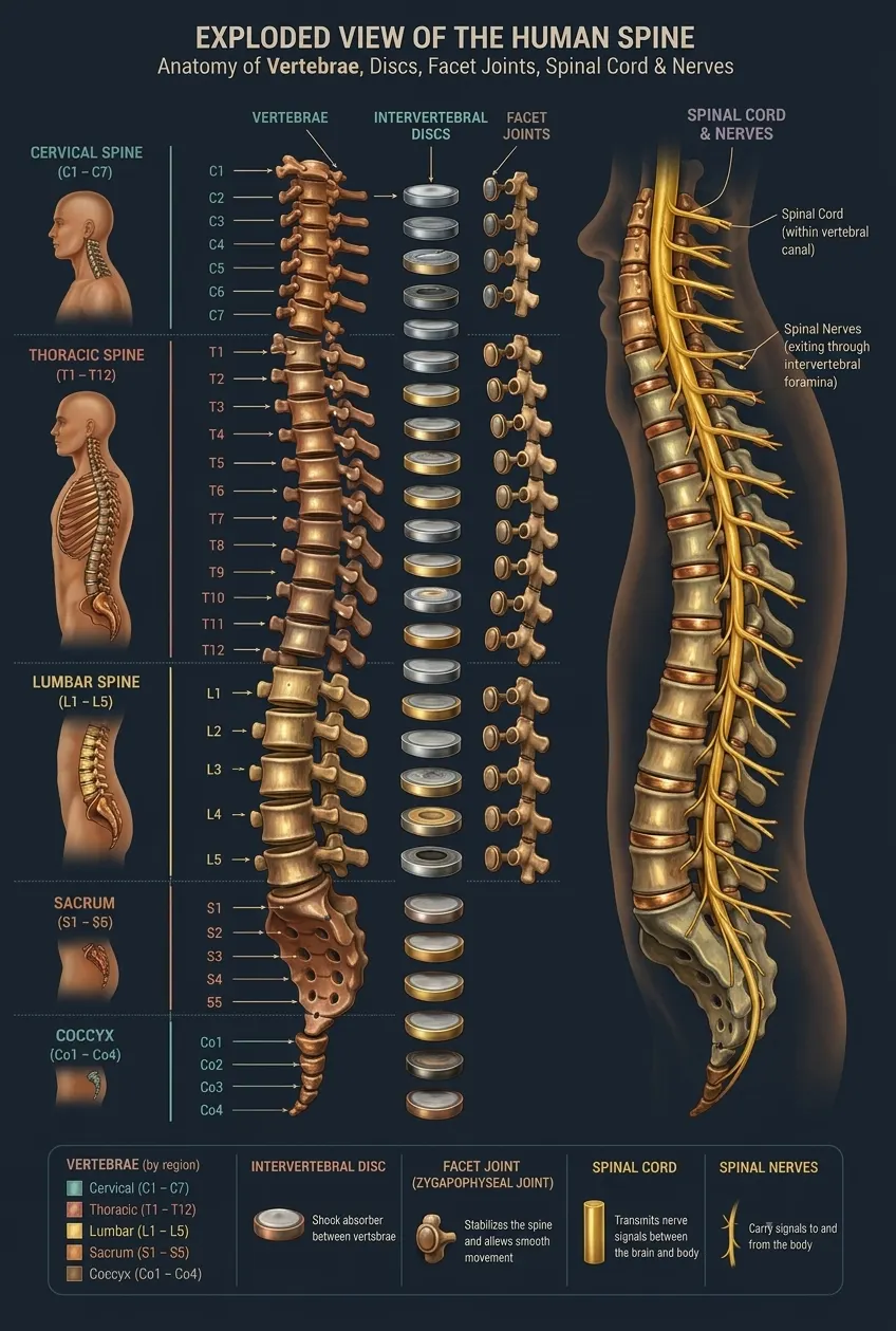 Enlarge image: Detailed anatomy of the human spine
Vertebrae and Facet Joints
Each vertebra has a strong vertebral body at the front and a bony arch behind it. The body is designed primarily for load-bearing, while the posterior arch helps form the spinal canal that surrounds the spinal cord and related neural structures.
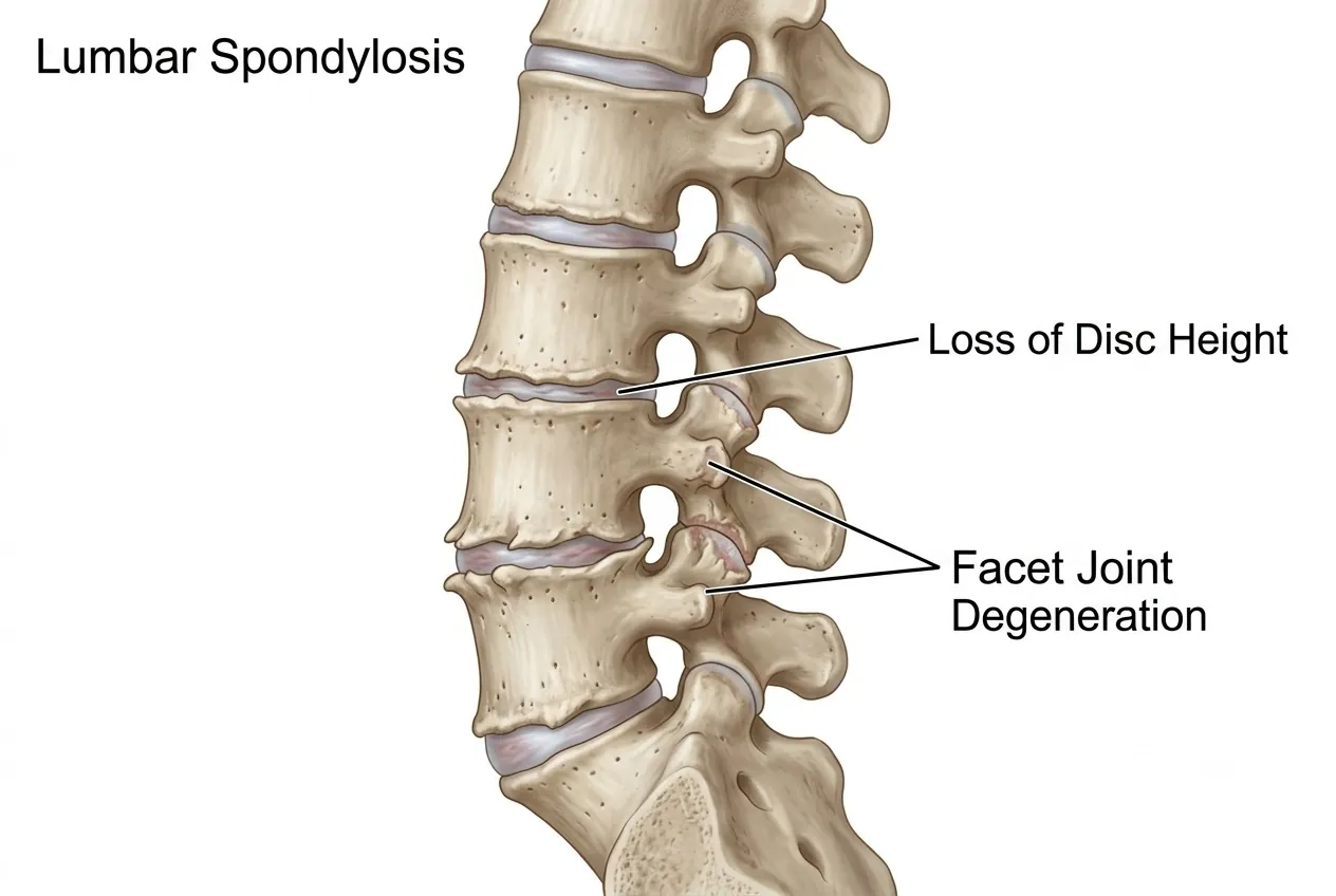
Neighbouring vertebrae are connected by intervertebral discs at the front and by paired facet joints at the back. Facet joints guide spinal movement and help prevent excessive or uncontrolled motion, allowing the spine to remain mobile without sacrificing stability.
Intervertebral Discs
Intervertebral discs are flexible cushioning structures located between most adjacent vertebral bodies. They help absorb impact, distribute pressure and permit controlled movement between vertebrae.
- Nucleus pulposus: the softer, gel-like central portion of the disc, which contributes to flexibility and shock absorption.
- Annulus fibrosus: the strong fibrous outer ring that surrounds the nucleus and helps maintain the shape and integrity of the disc.
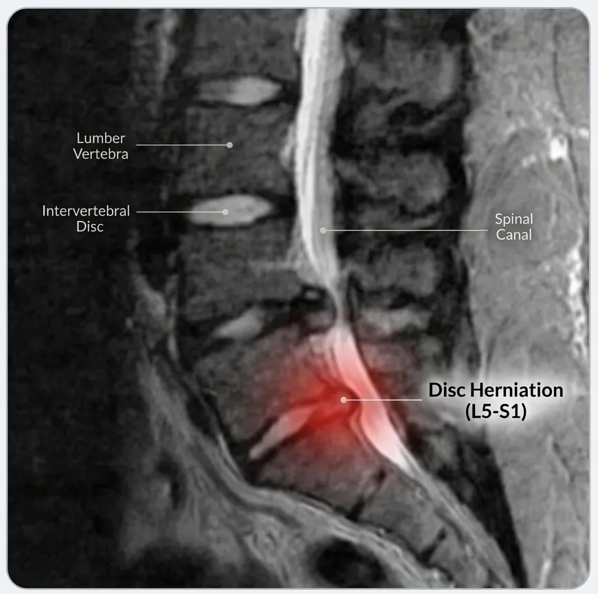
If the annulus develops a tear or weak area, part of the nucleus can move outward. This is commonly described as a disc herniation. When herniated disc material irritates or compresses a nearby nerve, symptoms can include radiating pain, numbness, tingling or weakness depending on the nerve involved.
Spinal Cord and Nerves
The spinal cord is the major communication pathway carrying signals between the brain and the rest of the body. It travels within the protected spinal canal, while spinal nerve roots branch away and leave the spine through openings called neural foramina.
There are 31 pairs of spinal nerves. Together, these nerves transmit sensory information and motor signals and contribute to the control of different regions and functions of the body.
- Cervical nerves: primarily supply areas including the neck, upper chest, shoulders and arms.
- Thoracic nerves: mainly supply the chest and abdominal wall.
- Lumbar and lower spinal nerves: contribute to sensation and movement in the legs and are associated with important lower-body functions, including pathways involved in bowel and bladder control.
Pressure on a nerve root may cause pain that travels into an arm or leg, along with numbness or weakness. Compression affecting the spinal cord can produce broader neurological problems such as difficulty with balance, hand coordination or walking and requires appropriate clinical assessment.
Why Spinal Anatomy Matters
The normal spine is designed to provide support, movement and protection at the same time. Its vertebrae carry load, discs cushion movement, facet joints guide motion, natural curves maintain balance, and the spinal canal protects the spinal cord and nerves.
Understanding this normal anatomy provides the foundation for recognising conditions such as disc herniation, nerve compression, abnormal spinal curvature and degenerative changes. Maintaining good posture, regular appropriate exercise and timely medical evaluation when persistent or neurological symptoms occur can help support long-term spinal health.
Imaging from Dr. Sharma's practice
The images below are from Dr. Amit Sharma's practice and are shared for education, without identifying details. Every patient's imaging is different — your own scans need an individual clinical review.
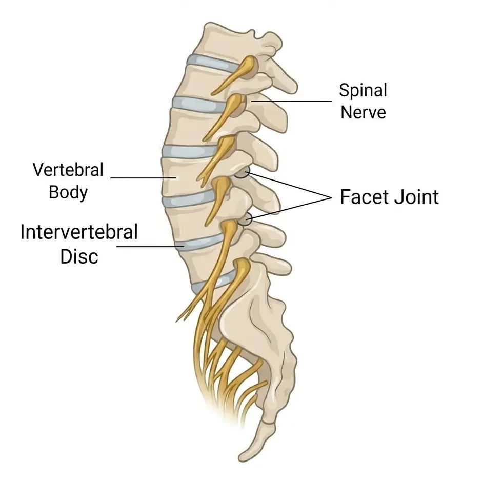 Enlarge image: Lumbar spine segment showing the vertebral body, intervertebral disc and spinal nerve.
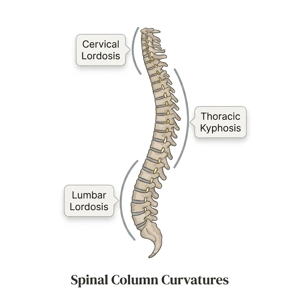 Enlarge image: The natural curves of the spine: cervical lordosis, thoracic kyphosis and lumbar lordosis.
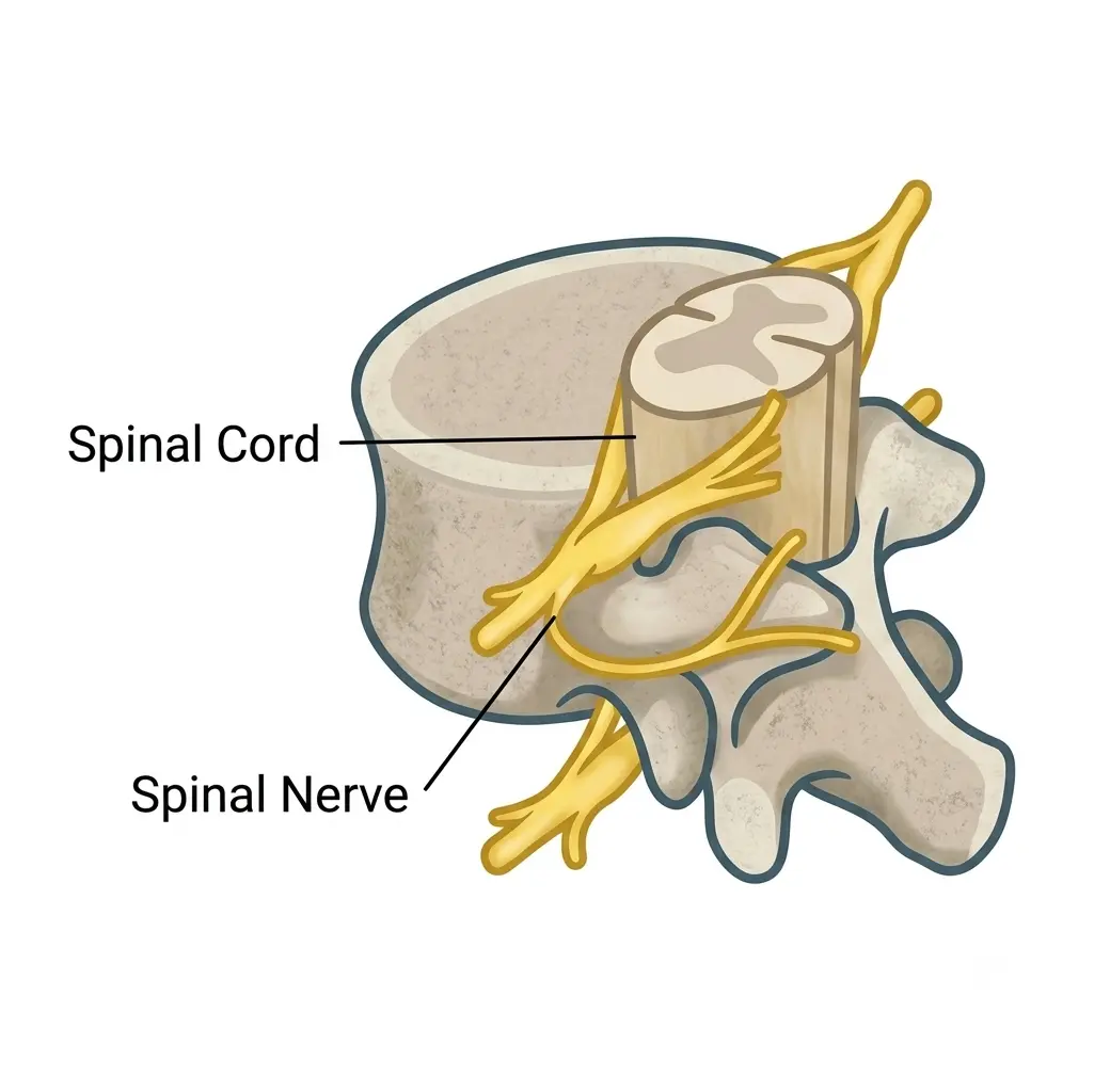 Enlarge image: A single spinal segment showing the vertebra, spinal cord and exiting spinal nerve.
This article is general education written by Dr. Amit Sharma. It is not a diagnosis or a substitute for an in-person consultation. If you have sudden weakness, numbness in the groin or saddle area, or new bladder or bowel problems, seek emergency care.