Start With the Impression
An MRI report can look alarming because radiologists describe every visible structural change, including findings that may be common with age and may not be responsible for pain. The most useful place to begin is the Impression or Conclusion near the end of the report. It summarizes the findings the radiologist considers most important.
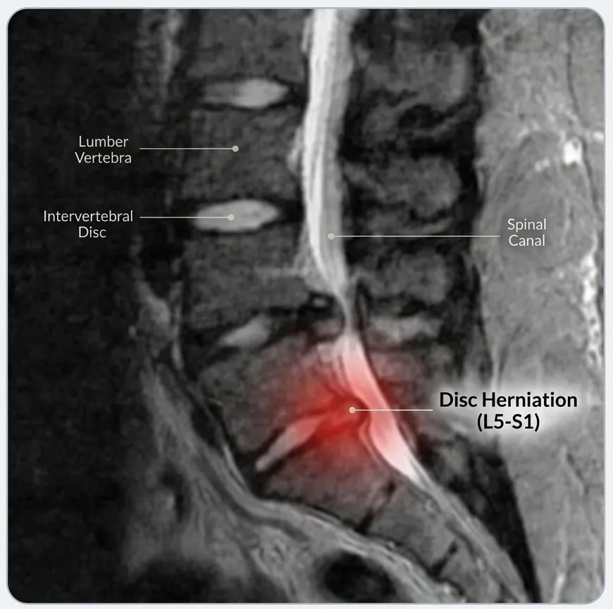
Next, identify the spinal level involved—such as C5–C6 in the neck or L4–L5 and L5–S1 in the lower back—and whether the report describes pressure on a nerve root, narrowing around the spinal cord, or simply age-related disc changes.
The key principle
An MRI is an anatomical picture, not a diagnosis by itself. Treatment decisions should be based on whether the scan findings match the patient's symptoms and neurological examination.
Common MRI Terms
Most spine MRI reports repeatedly use a small group of technical terms. Understanding them makes the report much easier to read.
| MRI term | What it generally means |
|---|---|
| Disc desiccation | Loss of water content in a disc; a common degenerative change. |
| Disc height loss | The disc has become thinner, usually from degeneration. |
| Osteophyte | A bony spur that develops around a degenerating joint or disc. |
| Facet arthropathy | Wear-and-tear change in the small joints at the back of the spine. |
| Ligamentum flavum hypertrophy | Thickening of a spinal ligament that can contribute to canal narrowing. |
| Canal stenosis | Narrowing of the central spinal canal. |
| Foraminal narrowing | Narrowing of the opening through which a spinal nerve exits. |
| Thecal sac indentation | A structure is touching or indenting the sac around the nerves; this does not automatically mean nerve injury. |
Disc Bulge, Protrusion and Extrusion
These words describe the shape and extent of disc displacement. They do not, by themselves, determine how severe a patient's condition is.
| Term | Practical meaning |
|---|---|
| Disc bulge | A broad extension of the disc beyond its usual boundary. |
| Protrusion | A more focal herniation in which the displaced portion remains relatively contained. |
| Extrusion | Disc material extends farther out through the outer disc covering. |
| Sequestration | A fragment of disc material has separated from the parent disc. |
A large-looking herniation can sometimes cause surprisingly little trouble, while a smaller one in a tight location can significantly irritate a nerve. Location and clinical correlation matter as much as size.
What Does Nerve Compression Mean?
Reports may use words such as contact, abutment, impingement or compression. These terms suggest increasing interaction between a disc, bone or ligament and a neural structure, but the wording still has to be interpreted alongside symptoms.
- Nerve-root compression can produce radiating arm or leg pain, tingling, numbness or weakness in the distribution of that nerve.
- Spinal-cord compression in the cervical or thoracic spine is more important when accompanied by hand clumsiness, balance problems, gait change, stiffness or other signs of myelopathy.
- Cauda equina compression in the lower spine becomes an emergency when associated with new bladder/bowel dysfunction, saddle numbness or rapidly progressive weakness.
Incidental and Age-Related Findings
Degenerative changes are frequently visible on MRI even in people who are functioning well. Disc dehydration, small bulges, mild facet arthritis and minor narrowing can therefore be incidental findings rather than the true source of symptoms.
Do not treat the report alone
A phrase such as “disc bulge” or “degenerative changes” is not an automatic indication for injections or surgery. The important question is whether that finding explains the patient's actual pain pattern or neurological deficit.
Why the MRI Must Match Your Symptoms
A spine specialist reads the scan together with the history and examination. For example, an L5 nerve problem should produce a pattern of symptoms and examination findings compatible with that nerve. If the report highlights a level that does not fit the clinical picture, that MRI finding may not be the pain generator.
What the doctor correlates with the MRI
- Where pain travels and what movements or positions trigger it.
- Areas of numbness or altered sensation.
- Muscle strength and any objective weakness.
- Reflexes, gait, balance and hand function where relevant.
- Previous treatment and whether symptoms are improving, stable or worsening.
When MRI Findings Become More Important
MRI findings carry greater clinical weight when they show a structural problem that clearly matches persistent or progressive neurological symptoms.
Seek urgent assessment
New bladder or bowel dysfunction, saddle numbness, rapidly worsening weakness, or major deterioration in walking or balance.
Arrange timely review
Persistent radiating pain, progressive numbness or weakness, or symptoms that continue despite appropriate treatment.
Often routine
Mild degenerative findings without neurological loss can usually be interpreted in the context of symptoms rather than treated as an emergency.
Questions to Ask About Your MRI
Take these questions to your consultation
- Which MRI finding actually explains my symptoms?
- Is a nerve root or the spinal cord truly compressed?
- Are any of the reported changes incidental or age-related?
- Do I need treatment now, or can this be observed?
- What non-surgical options are appropriate?
- What warning signs should make me seek urgent care?
- If surgery is being considered, what specific anatomical problem would it correct?
The aim is not to decode every medical word in isolation. It is to understand which findings matter clinically and what they mean for the next step in care.
This article is general education written by Dr. Amit Sharma. It is not a diagnosis or a substitute for an in-person consultation.