Introduction
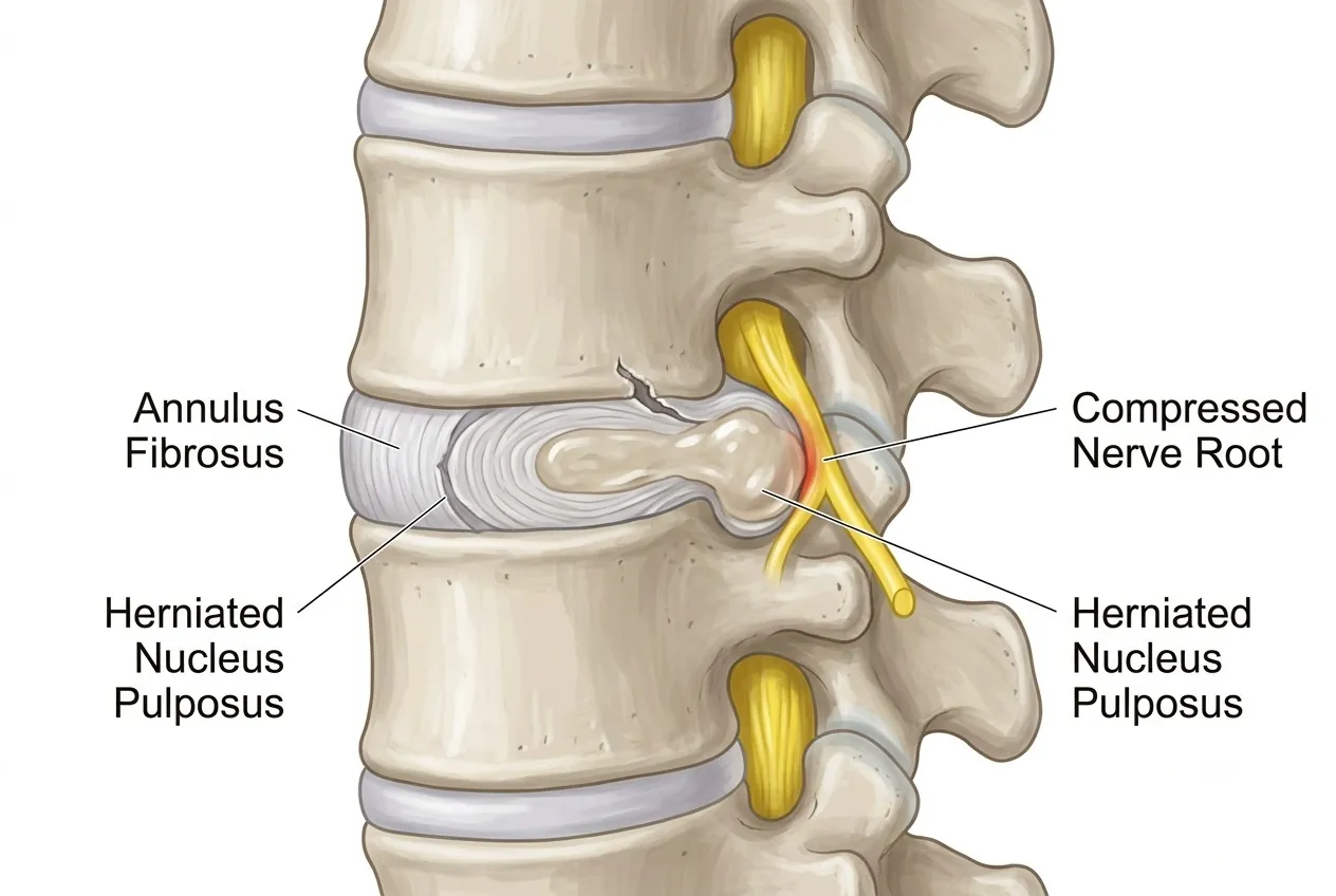
A slipped disc, more accurately called a herniated disc, is a common spinal condition in which part of the softer inner disc material pushes outward through a weakened or torn area of the tougher outer layer. The inner portion is called the nucleus pulposus, while the outer ring is the annulus fibrosus.
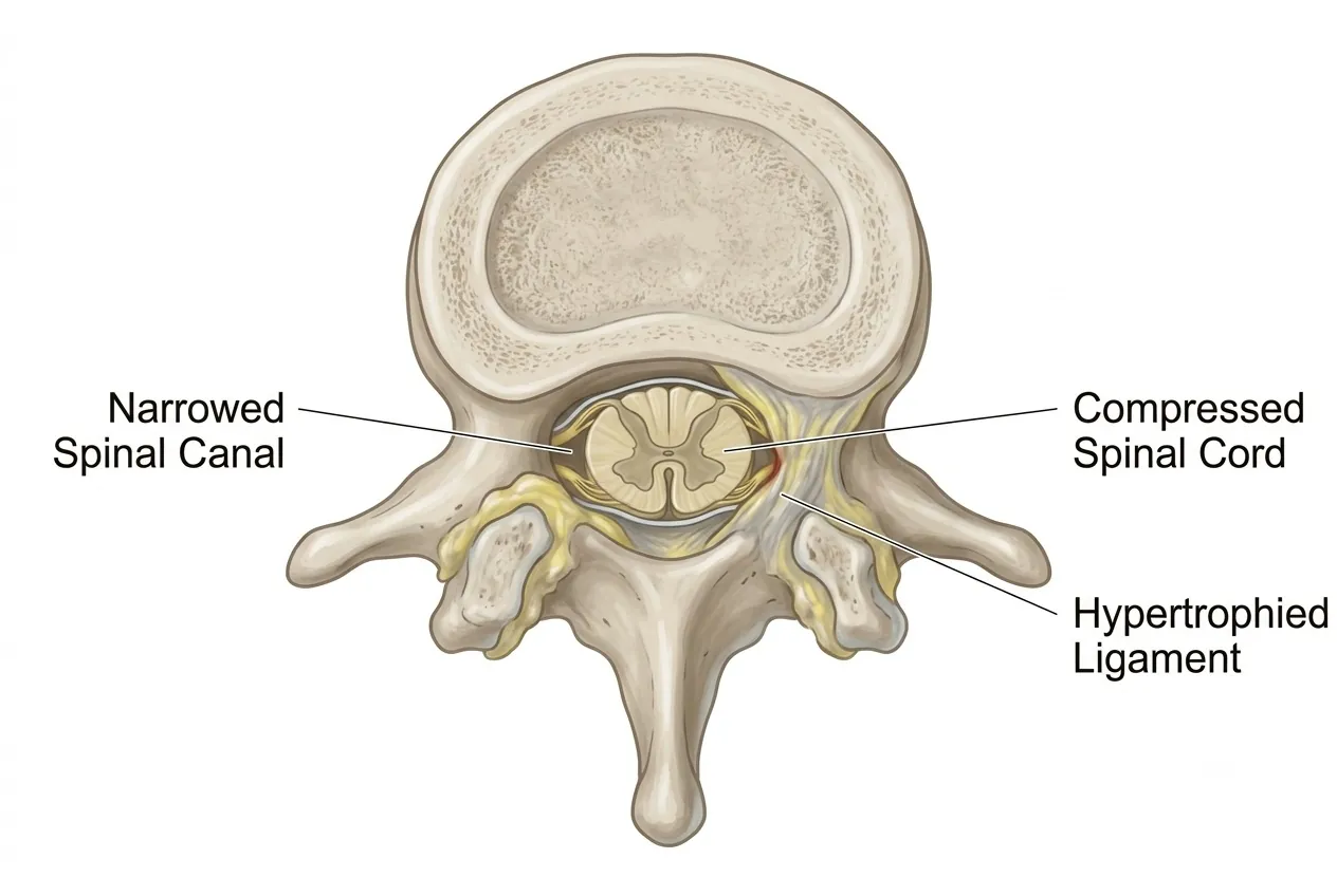
Disc herniation becomes more common as discs undergo age-related degeneration, but it can also affect younger adults. Symptoms develop when the damaged disc itself becomes painful or when displaced disc material irritates or compresses a nearby nerve root or, in some regions of the spine, the spinal cord.
A herniated disc can occur in the cervical, thoracic or lumbar spine. Lumbar disc herniation is particularly well known for causing sciatica, while a cervical disc herniation can produce pain, tingling, numbness or weakness extending into an arm.
Causes and Risk Factors
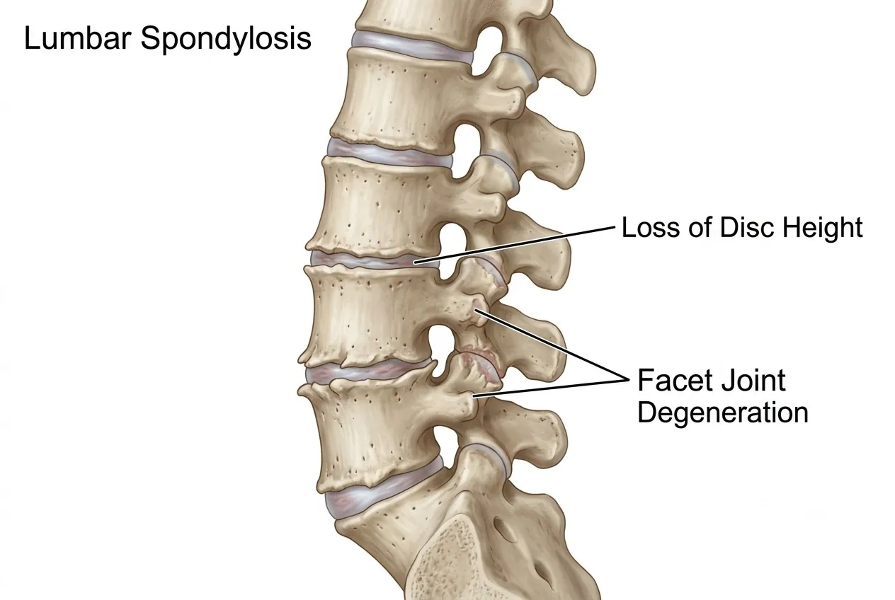
The most important underlying factor is degeneration of the intervertebral disc. With age, discs gradually lose water content, a process known as disc desiccation. A less hydrated disc is less elastic and may become more susceptible to fissuring, bulging or herniation.
- Prolonged poor posture and inactivity: sustained loading and weak supporting muscles can increase mechanical stress on the spine.
- Excess body weight: additional weight increases the load carried by spinal discs, particularly in the lower back.
- Trauma: falls, accidents and sudden injuries can damage a disc.
- Improper heavy lifting: lifting a substantial load while bending or twisting can place high stress on the lumbar discs.
- Genetic predisposition: inherited differences in connective tissue and disc degeneration can make some individuals more susceptible.
Types of Disc Herniation
MRI reports may use different terms to describe the shape and extent of displaced disc material. These descriptions are useful, but treatment is never decided from an MRI label alone. The scan must be interpreted together with the patient's symptoms and neurological examination.
Disc Bulge or Protrusion
- The outer disc contour extends beyond its usual boundary while the displaced material remains connected to the disc.
- Symptoms may be mild or absent unless the bulge irritates or compresses a nerve.
- Many symptomatic cases can be managed with medication, activity modification and physiotherapy.
Disc Extrusion
- Disc material extends through a defect in the annulus and may produce more substantial nerve or spinal-canal compression.
- Treatment depends on pain severity, neurological findings and the relationship between the MRI abnormality and symptoms.
- Management may range from conservative care to an epidural injection or surgery in selected patients.
Disc Sequestration
- A fragment of extruded disc material separates from the parent disc and lies within the spinal canal.
- A sequestrated fragment can cause marked nerve compression, although some fragments may reduce in size or resorb over time.
- The need for surgery depends on neurological deficit, pain, emergency symptoms and response to appropriate non-operative treatment rather than the MRI appearance alone.
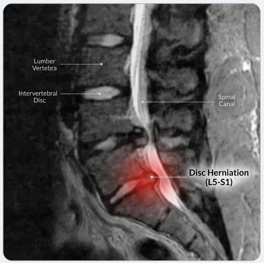
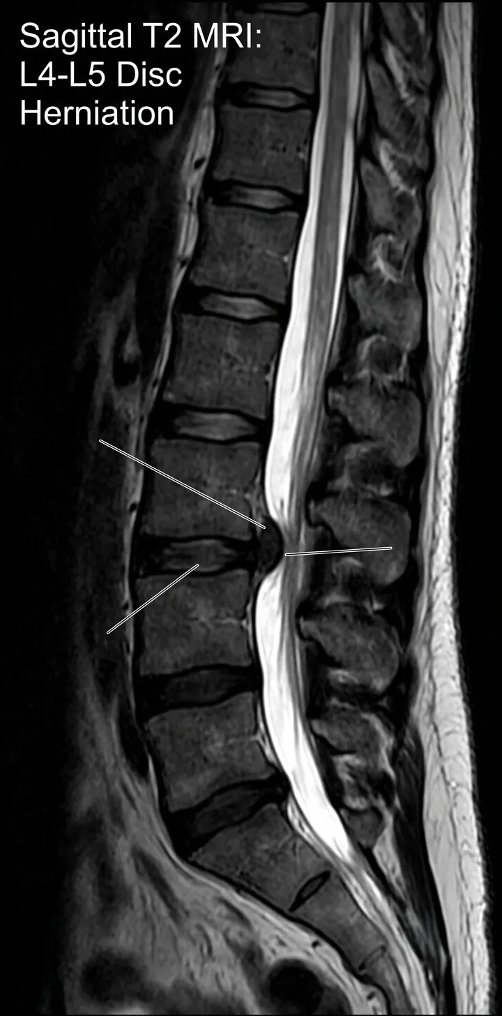 Enlarge image: MRI example of a slipped or herniated disc
Symptoms
Symptoms depend on the spinal level involved, the direction and size of the herniation and whether a nerve root or the spinal cord is compressed. Importantly, some disc bulges and herniations identified on imaging produce no symptoms.
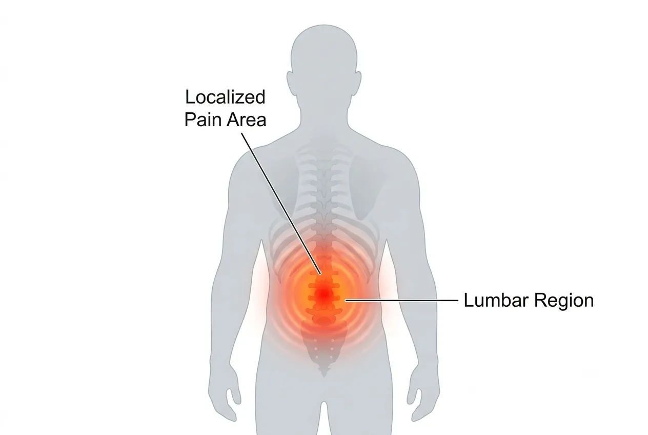
- Local pain: pain may occur in the neck with cervical disc disease, the upper or middle back with thoracic disease, or the lower back with lumbar disc disease.
- Radicular pain: nerve-root irritation can cause pain travelling into an arm or leg. Lumbar nerve compression commonly produces sciatica.
- Numbness or tingling: altered sensation may occur along the distribution of the affected nerve.
- Muscle weakness: significant nerve compression can reduce strength in particular muscle groups.
- Stiffness or balance difficulty: cervical or thoracic disease affecting the spinal cord may cause broader neurological symptoms.
- Bladder or bowel dysfunction: new loss of bladder or bowel control associated with severe spinal symptoms is an emergency warning sign and requires urgent medical assessment.
2-minute self-check
Do your symptoms deserve a closer look?
Tick anything you've noticed recently. Your guidance appears straight below. Everything stays on this page; nothing is stored or sent. This is an educational aid, not a diagnosis of slip disc.
Seek urgent medical care today
These symptoms can mean a nerve or the spinal cord is under serious pressure. Go to the nearest emergency department, or call the clinic straight away. Do not wait for a routine appointment.
Book a spine specialist review soon
Arrange an assessment within the next few days, and sooner if things get worse. If you already have an MRI, you can send it for review.
Conservative care is usually a reasonable start
Activity changes, physiotherapy and appropriate medication often help. See a doctor if it is not better in 1–2 weeks, or straight away if any time-sensitive symptom appears.
Guidance only, based on the warning signs described in this guide. It cannot find the cause of your symptoms. If symptoms are severe, getting worse quickly, or you are worried, contact a doctor.
Diagnosis
Diagnosis begins with a detailed history and physical examination. The pattern of pain, numbness and weakness is compared with muscle strength, sensation, reflexes and nerve-tension signs to determine whether a particular nerve is likely to be involved.
Investigations Used for a Suspected Slip Disc
- X-ray: does not directly show a herniated disc but can help assess alignment, degeneration and other bony abnormalities.
- MRI: is the principal imaging investigation for visualising discs, nerve roots, the spinal canal and the extent of a herniation.
- EMG/NCV: may be used in selected cases when further assessment of nerve function is necessary.
- CT scan: can provide detailed information about bony anatomy and may be useful when MRI cannot be performed or when additional bone detail is required.
An MRI abnormality must correlate with the clinical problem. A disc bulge seen on a scan does not automatically require treatment or surgery if it is not responsible for the patient's symptoms.
Non-Surgical Treatment
Many patients with a symptomatic herniated disc improve without surgery. Treatment aims to control pain, allow the irritated nerve to settle and gradually restore normal movement and strength.
- Medication: depending on the patient, treatment may include appropriate anti-inflammatory or other analgesic medication, short-term medication for muscle spasm, and neuropathic-pain medicines such as gabapentin or pregabalin when nerve pain is present.
- Controlled activity: a brief reduction in painful activity may help during an acute episode, but prolonged complete bed rest or immobilisation is generally avoided.
- Physiotherapy: rehabilitation can address movement, flexibility, posture, core control and progressive strengthening.
- Home exercise: stretching and strengthening may form part of recovery, but the appropriate exercise depends on the level of the herniation and the patient's symptoms and should preferably be selected with professional guidance.
- Ergonomic and lifestyle measures: sensible lifting technique, posture, weight management and appropriate sleep support can reduce unnecessary spinal loading.
Epidural Injections
For selected patients with significant nerve-related pain, an epidural steroid or targeted nerve-root injection may be considered. The medication is placed close to the irritated neural structure to reduce inflammation and provide pain relief while natural recovery and rehabilitation continue.
An injection may be useful when pain is severe despite medication, when temporary relief is needed to allow rehabilitation, when surgery needs to be postponed or is not presently desired, or in selected multi-level cases where a targeted injection may help clarify which level is producing symptoms. Relief can be temporary, and injections do not mechanically remove the herniated disc.
Surgical Treatment
Surgery is considered when there is a clear clinical indication—for example persistent disabling nerve pain despite appropriate conservative treatment, progressive neurological weakness, significant spinal-cord compression or an emergency neurological syndrome. The operation chosen depends on the spinal level, anatomy, degree of compression and presence of instability or degeneration.
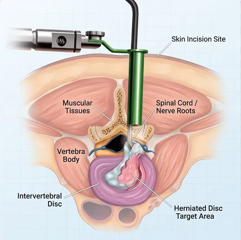
- Microdiscectomy: the herniated portion compressing a nerve is removed through a small surgical approach, commonly using magnification or a microscope.
- Endoscopic discectomy: selected disc herniations can be treated through an endoscopic approach designed to minimise tissue disruption.
- Other minimally invasive disc procedures: certain carefully selected contained disc problems may be considered for procedures intended to reduce disc pressure; suitability depends strongly on the exact pathology.
- Spinal fusion: fusion may be required in selected cases involving instability, substantial degeneration, recurrent herniation or another structural reason that makes decompression alone insufficient.
The presence of a disc herniation on MRI is not, by itself, an indication for surgery. The decision is based on symptoms, neurological findings, imaging correlation and the patient's response to non-surgical care.
Recovery and Prevention
A slipped disc is common and often manageable. Recovery generally involves controlling the acute symptoms and then progressively returning to movement, conditioning and normal activity. Complete immobilisation for prolonged periods can contribute to stiffness and deconditioning.
Signs of improvement can include decreasing radiating pain, improved ability to sit or walk, reduced numbness or tingling and gradual return of strength. Recovery time varies considerably according to the severity and location of the herniation.
- Use appropriate lifting technique and avoid combining heavy lifting with uncontrolled twisting.
- Maintain regular physical activity and develop trunk and spinal-supporting strength.
- Address prolonged poor posture and improve workstation ergonomics where necessary.
- Maintain a healthy body weight to reduce excessive spinal loading.
- Increase walking and other activity gradually according to symptoms and professional advice.
Imaging from Dr. Sharma's practice
The images below are from Dr. Amit Sharma's practice and are shared for education, without identifying details. Every patient's imaging is different — your own scans need an individual clinical review.
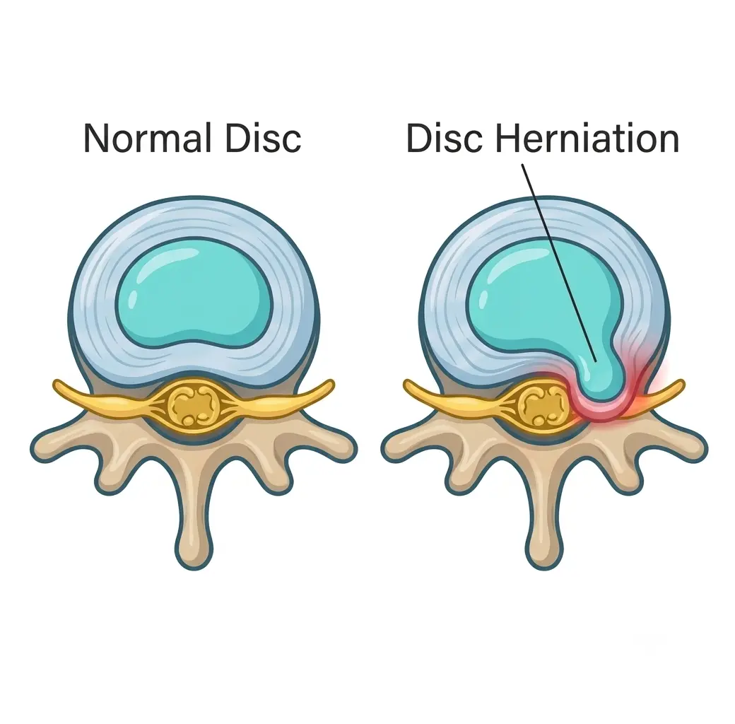 Enlarge image: Illustration comparing a normal lumbar disc with a disc herniation (lumbar disc prolapse) pressing on the nerve.
Frequently Asked Questions
How can a herniated disc heal as quickly as possible?
There is no instant cure. Early treatment usually combines sensible activity modification, pain control and gradual rehabilitation. Complete prolonged bed rest should generally be avoided. Physiotherapy, appropriate core and mobility exercises, ergonomic improvements and, in selected cases, an epidural injection can support recovery.
What are signs that a herniated disc is improving?
Improvement may include less arm or leg pain, better tolerance of walking and daily activity, reduced tingling or numbness and recovery of muscle strength. Persistent or worsening weakness should be reassessed rather than simply observed.
Is walking good for a herniated disc?
Walking is often a useful low-impact activity when tolerated. Begin at a comfortable duration and increase gradually. Severe worsening of radiating pain, weakness or neurological symptoms warrants reassessment.
Which exercises should be avoided?
There is no universal prohibited-exercise list because the correct movement strategy depends on the location and behaviour of the disc problem. During an acute painful phase, avoid movements or heavy loading that repeatedly reproduce or intensify radiating symptoms, and obtain guidance before beginning aggressive strengthening.
Can a slipped or bulging disc be treated without surgery?
Yes. Many patients improve with medication, controlled activity, physiotherapy and time. In selected cases, injections may help. Surgery is reserved for specific indications such as persistent disabling nerve pain, progressive neurological deficit or emergency compression.
What is the best painkiller for a herniated disc?
The appropriate medication depends on whether the dominant pain is inflammatory, muscular or neuropathic and on the patient's other medical conditions. Anti-inflammatory medicines, other analgesics, muscle relaxants or neuropathic-pain medicines may be used selectively. Medication should be chosen with a clinician rather than assuming one drug is best for every patient.
What are the emergency symptoms of a herniated disc?
Seek urgent medical assessment for new bladder or bowel dysfunction, numbness around the groin or saddle region, rapidly progressive leg weakness, severe neurological deterioration or symptoms suggesting major spinal-cord compression. These can indicate serious nerve compression requiring prompt evaluation.
This article is general education written by Dr. Amit Sharma. It is not a diagnosis or a substitute for an in-person consultation. If you have sudden weakness, numbness in the groin or saddle area, or new bladder or bowel problems, seek emergency care.