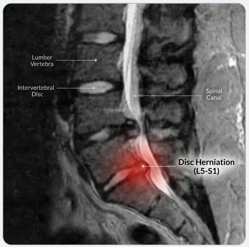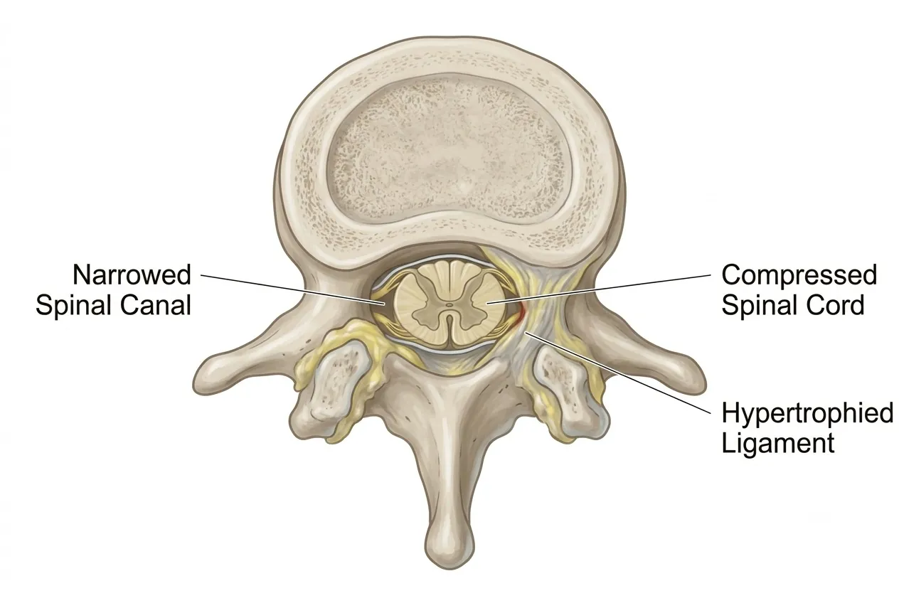
What Is Spinal Stenosis?
Spinal stenosis means narrowing of the spinal canal or the smaller passages through which spinal nerves travel. As these spaces become tighter, the spinal cord or nerve roots may be compressed, producing pain, numbness, weakness, walking difficulty or other neurological symptoms.
Some people are born with a relatively narrow spinal canal, known as congenital or developmental stenosis. In these patients, even modest age-related changes may eventually reduce the available space enough to cause symptoms. More commonly, stenosis develops gradually as discs, joints, ligaments and bones undergo degenerative change.
The MRI alone does not define the problem
The degree of narrowing seen on a scan must be interpreted together with symptoms and neurological examination. Some patients have significant-looking stenosis with limited symptoms, while others are functionally restricted by nerve compression.
Causes of Spinal Stenosis
Stenosis usually develops because several structures progressively reduce the space available for the nerves rather than because of a single abnormality.
Common Degenerative Causes
- Disc bulging or herniation: displaced disc tissue can encroach on the central canal or nerve-root passage.
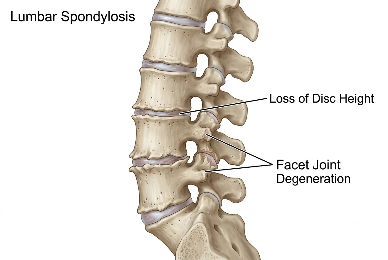
- Facet-joint arthritis: enlarged, arthritic joints can progressively reduce space around the nerves.
- Ligamentum flavum hypertrophy: this ligament can thicken and buckle inward as degeneration progresses.
- Loss of disc height: disc collapse can narrow the neural foramina through which spinal nerves exit.
- Age-related degeneration: combinations of disc wear, bone overgrowth and joint change frequently contribute.
Other Causes and Contributing Factors
- Developmental narrowing: a naturally smaller spinal canal leaves less reserve space for later degenerative change.
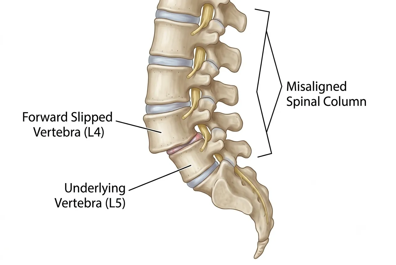
- Spondylolisthesis: slippage of one vertebra relative to another can narrow the canal and foramina.
- Fracture or spinal injury: altered anatomy or displaced bone can compromise neural space.
- Infection or tuberculosis: destructive or inflammatory spinal disease can produce secondary stenosis.
- Spinal tumour: a mass within or around the spinal canal can compress neural structures.
- Mechanical factors: obesity, heavy physical work and poor spinal mechanics may contribute to degenerative stress, although they are not the sole cause of stenosis.
Symptoms by Spinal Region
The clinical pattern depends strongly on where the narrowing occurs. Lumbar stenosis mainly affects the legs, while cervical and thoracic stenosis can involve the spinal cord and therefore produce broader neurological problems.
| Region | Typical Symptoms | Important Pattern |
|---|---|---|
| Lumbar | Leg pain, tingling, numbness, heaviness, weakness or reduced sensation. | Symptoms often appear after standing or walking and improve with sitting or bending forward — a pattern called neurogenic claudication. |
| Thoracic | Walking difficulty, imbalance, leg stiffness or spasticity, with symptoms that may resemble lumbar disease. | Thoracic cord compression can produce myelopathy and should be identified before permanent neurological impairment develops. |
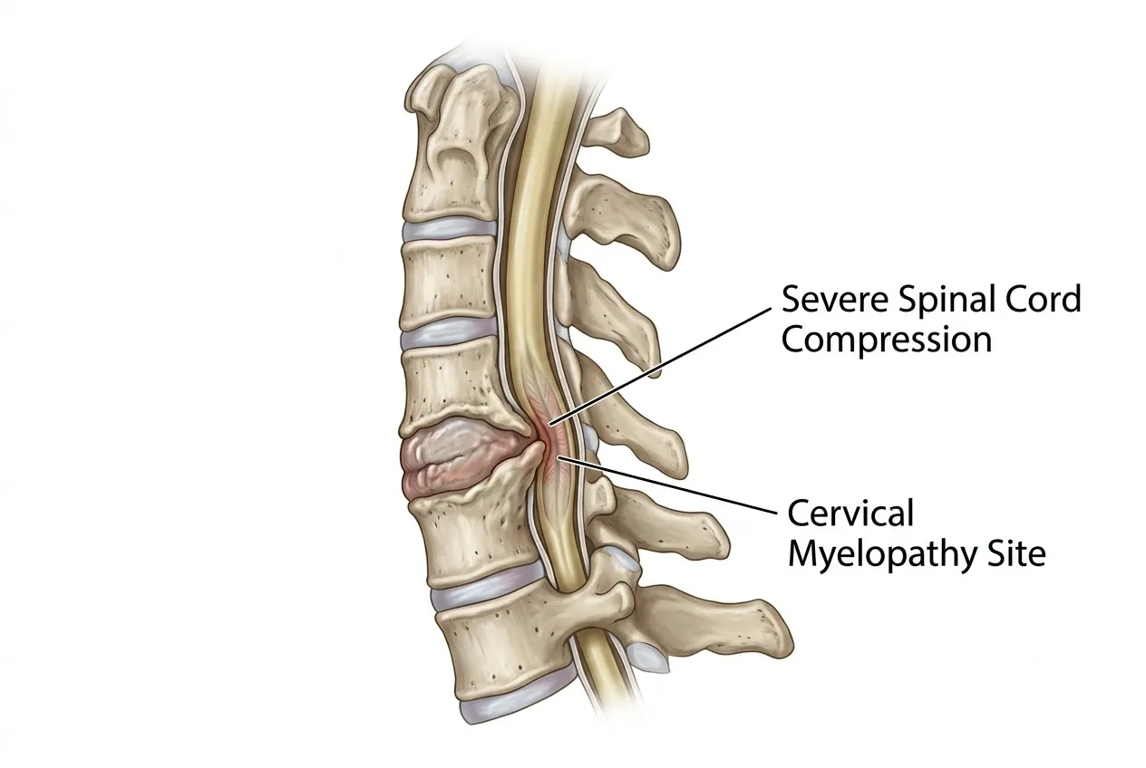
| Cervical | Hand numbness, weakness, reduced grip strength, loss of dexterity and arm symptoms. | More severe cord compression may cause cervical myelopathy with gait imbalance, limb stiffness and, in advanced cases, bladder or bowel dysfunction. |
Neurogenic claudication can resemble vascular claudication caused by reduced circulation to the legs. A detailed clinical assessment, and sometimes vascular testing, helps distinguish between these conditions.
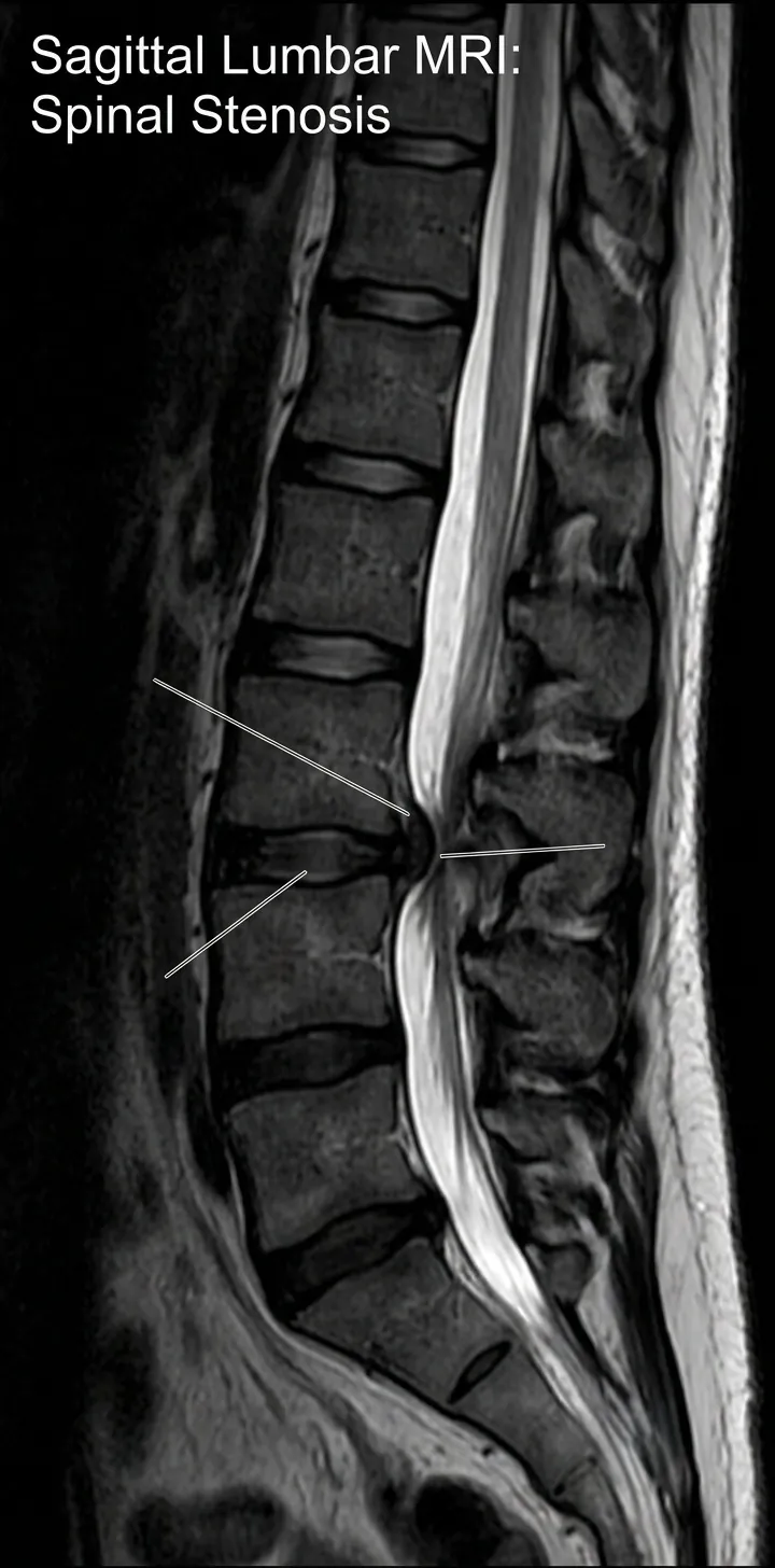 Enlarge image: MRI showing spinal canal stenosis
2-minute self-check
Do your symptoms deserve a closer look?
Tick anything you've noticed recently. Your guidance appears straight below. Everything stays on this page; nothing is stored or sent. This is an educational aid, not a diagnosis of spinal stenosis.
Seek urgent medical care today
These symptoms can mean a nerve or the spinal cord is under serious pressure. Go to the nearest emergency department, or call the clinic straight away. Do not wait for a routine appointment.
Book a spine specialist review soon
Arrange an assessment within the next few days, and sooner if things get worse. If you already have an MRI, you can send it for review.
Conservative care is usually a reasonable start
Activity changes, physiotherapy and appropriate medication often help. See a doctor if it is not better in 1–2 weeks, or straight away if any time-sensitive symptom appears.
Guidance only, based on the warning signs described in this guide. It cannot find the cause of your symptoms. If symptoms are severe, getting worse quickly, or you are worried, contact a doctor.
Diagnosis
Diagnosis combines the patient's symptoms with neurological examination and appropriate imaging. The purpose is not only to demonstrate narrowing but also to determine whether it explains the functional or neurological problem.
Investigations for Spinal Stenosis
- Medical history and neurological examination: assesses walking, strength, sensation, reflexes and signs of nerve-root or spinal-cord dysfunction.
- MRI spine: provides detailed assessment of the canal, discs, ligaments, nerve roots and spinal cord and is the principal imaging investigation for stenosis.
- X-rays: demonstrate alignment, degeneration, vertebral slippage and other bony changes; selected views may help assess instability.
- CT scan: provides detailed bony anatomy and can be useful when MRI is contraindicated or additional bone detail is required.
- EMG/NCV: may help assess nerve function when symptoms and imaging do not clearly correspond.
- Doppler study: may be requested when vascular claudication needs to be excluded.
- Blood tests: may help investigate alternative or contributing conditions such as vitamin deficiency, infection or systemic disease.
- Biopsy and histopathology: may be necessary when a tumour or infection is suspected.
Non-Surgical Treatment
Patients with manageable symptoms and no progressive neurological deficit can often begin with non-operative treatment. The goal is to improve function and control symptoms; treatment does not necessarily reverse the structural narrowing itself.
- Medication: appropriate analgesic or anti-inflammatory treatment may reduce pain sufficiently to maintain activity.
- Activity modification: pacing activities and avoiding repeatedly provocative positions can improve day-to-day function.
- Physiotherapy: treatment may focus on mobility, conditioning, trunk strength, posture and strategies that improve walking tolerance.
- Weight and general health management: reducing unnecessary mechanical load and maintaining physical conditioning can support symptom control.
Epidural steroid injections may provide temporary relief in selected patients by reducing inflammation around irritated nerves. The duration of benefit varies; an injection does not permanently enlarge a narrowed spinal canal. Suitability also depends on factors such as diabetes, anticoagulant or antiplatelet medication, infection risk and the patient's overall medical condition.
When to Seek Medical Care
The urgency of assessment depends on the symptoms. Progressive neurological problems deserve more attention than pain alone.
Seek urgent assessment
New bladder or bowel dysfunction, rapidly progressive weakness, severe deterioration in walking, or major symptoms suggesting spinal-cord or cauda-equina compression require prompt medical evaluation.
Arrange specialist review
Increasing walking limitation, persistent leg or arm numbness, hand clumsiness, balance problems or weakness should be assessed rather than repeatedly treated as simple back or neck pain.
Routine conservative care
Stable, manageable symptoms without neurological progression can often begin with medication, physiotherapy, activity modification and clinical follow-up.
Surgical Treatment
Surgery is considered when conservative care no longer provides acceptable function, walking becomes severely restricted, neurological deficits progress, or spinal-cord compression creates a risk of further neurological damage. The central goal is decompression: creating adequate space for the affected nerves or spinal cord.
| Procedure | How It Is Used |
|---|---|
| Micro-lumbar decompression | Uses a limited surgical approach and magnification to remove structures compressing lumbar nerves while minimising unnecessary tissue disruption. |
| Endoscopic decompression | In selected cases, an endoscope can be used through a small access route to decompress neural structures. Suitability depends on the stenosis pattern and patient anatomy. |
| Laminectomy | Removes part of the posterior vertebral bone to enlarge the spinal canal, particularly when more extensive or multilevel decompression is required. |
| Fusion / instrumentation | May be combined with decompression when clinically significant instability, spondylolisthesis, deformity or severe degeneration means decompression alone may not provide adequate stability. |
| MIS TLIF / Endoscopic-assisted fusion | Minimally invasive fusion techniques may be considered in selected lumbar stenosis cases that also require stabilisation. |
Not every stenosis patient needs fusion
Decompression alone may be sufficient when there is no clinically important instability. Fusion is added when the underlying structural problem requires stabilisation.
Imaging from Dr. Sharma's practice
The images below are from Dr. Amit Sharma's practice and are shared for education, without identifying details. Every patient's imaging is different — your own scans need an individual clinical review.
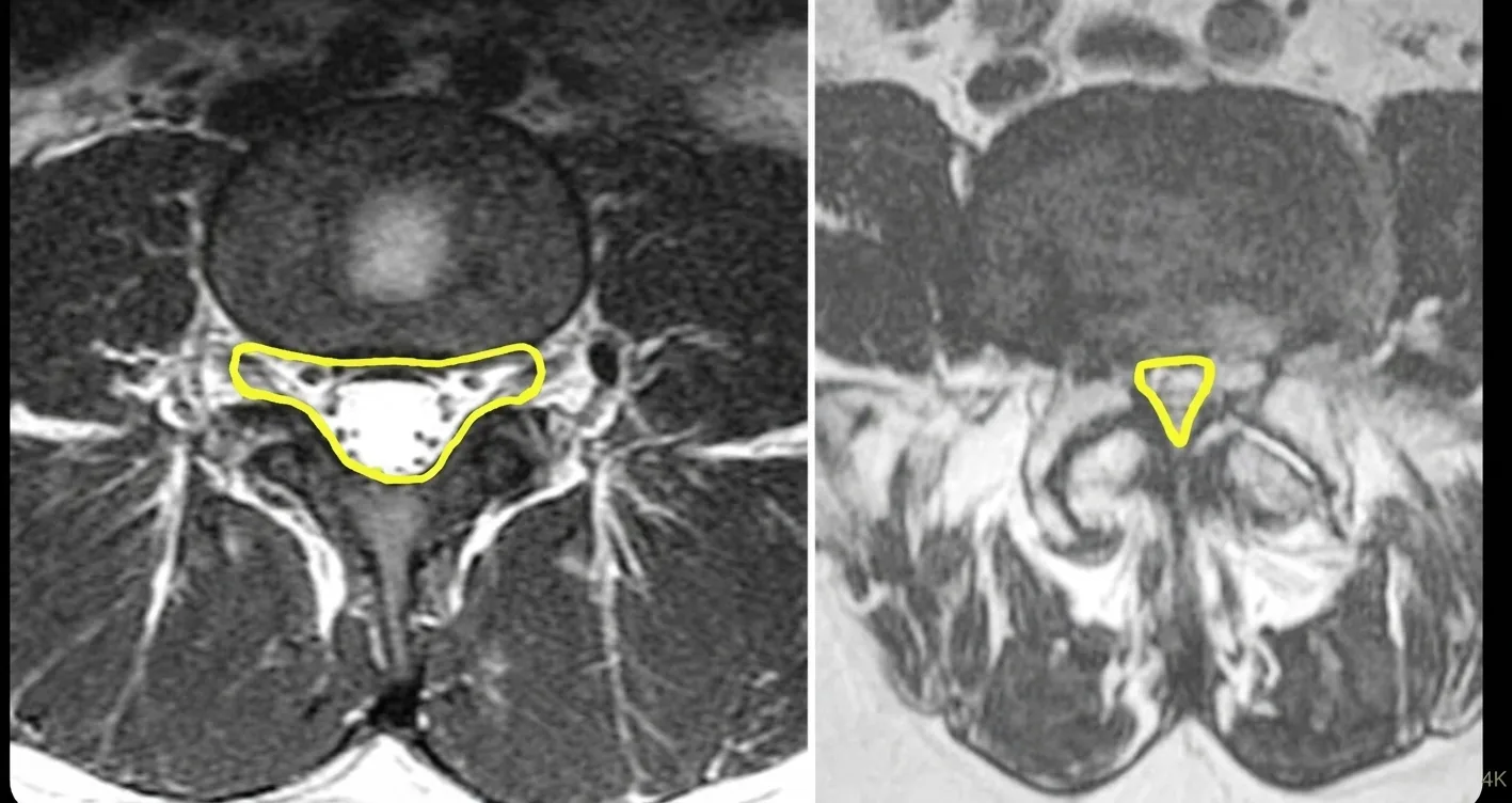 Enlarge image: Axial lumbar MRI: the outlined spinal canal is roomy on the left and markedly narrowed on the right.
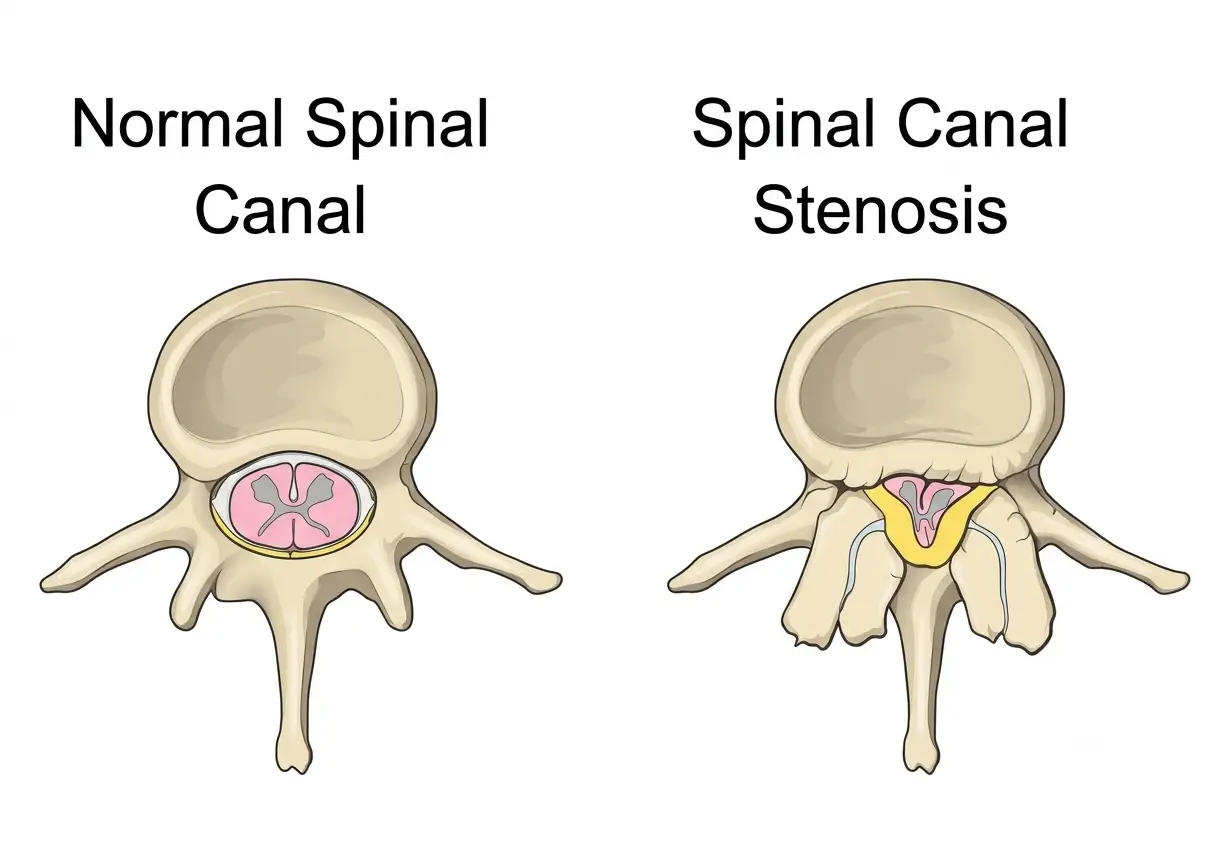 Enlarge image: Illustration comparing a normal spinal canal with spinal canal stenosis.
Frequently Asked Questions
What are the common causes of spinal stenosis?
Age-related degeneration is the most common cause. Disc bulging, facet-joint arthritis, ligamentum flavum thickening, disc-height loss and spondylolisthesis can all reduce neural space. Developmental narrowing, tumours, fractures and infections are less common causes.
Can spinal stenosis be treated without surgery?
Yes. Many patients with moderate, stable symptoms can be managed with medication, physiotherapy, activity modification and selected injections. Surgery becomes more relevant when function remains poor or neurological problems progress.
When should surgery be considered?
Surgery may be considered for severe walking limitation, persistent disabling nerve symptoms despite appropriate treatment, progressive weakness, spinal-cord dysfunction or other significant neurological compression.
What surgical options are available?
Depending on the anatomy, options include limited decompression, endoscopic decompression, laminectomy and decompression combined with fusion or instrumentation when instability is present.
How successful is minimally invasive surgery?
Outcome depends on the diagnosis, severity of nerve damage, procedure selected and overall patient health. Minimally invasive access can reduce tissue disruption in appropriately selected cases, but it does not guarantee a particular outcome.
How long is recovery after stenosis surgery?
Recovery varies with the procedure. A limited decompression and a fusion have different recovery timelines, and age, neurological status and other medical conditions also influence rehabilitation.
What does spinal stenosis surgery cost in Mumbai?
Cost varies according to the spinal level, number of levels, hospital, procedure, implants if required and length of stay. A meaningful estimate requires evaluation and a defined treatment plan.
What is the role of physiotherapy after surgery?
Rehabilitation can help restore walking tolerance, mobility, strength and confidence after the surgeon determines that exercise progression is appropriate.
Is spinal stenosis curable?
Degenerative narrowing cannot always be reversed without physically decompressing the affected area, but symptoms can often be controlled without surgery. When surgery is required, its purpose is to relieve neural compression and improve function.
How does ligamentum flavum hypertrophy cause stenosis?
The ligamentum flavum lies along the back of the spinal canal. With degeneration it can thicken or buckle inward, reducing the space available for the nerves.
Why does lumbar stenosis cause walking problems?
Standing and walking can reduce the functional space available to already compressed lumbar nerves, producing pain, heaviness, numbness or weakness. Sitting or bending forward often relieves this neurogenic claudication pattern.
What is the best painkiller for spinal stenosis?
There is no single best medication for every patient. Choice depends on pain type, age, kidney and stomach health, other medicines and medical conditions. Medication should be individualised by the treating clinician.
Which exercises should be avoided?
Avoid exercises that repeatedly worsen neurological symptoms or provoke significant leg or arm pain. The appropriate programme depends on the region and severity of stenosis and is best tailored by a clinician or physiotherapist.
Can spinal stenosis be fatal?
Degenerative spinal stenosis itself is not usually a life-threatening condition. Severe compression can, however, cause major and sometimes permanent neurological disability, which is why progressive weakness, bladder or bowel changes and spinal-cord symptoms require urgent assessment.
This article is general education written by Dr. Amit Sharma. It is not a diagnosis or a substitute for an in-person consultation. If you have sudden weakness, numbness in the groin or saddle area, or new bladder or bowel problems, seek emergency care.